• 文献检索
  • 文档翻译
  • 深度研究
  • 学术资讯
  • Suppr Zotero 插件Zotero 插件
  • 邀请有礼
  • 套餐&价格
  • 历史记录
应用&插件
Suppr Zotero 插件Zotero 插件浏览器插件Mac 客户端Windows 客户端微信小程序
定价
高级版会员购买积分包购买API积分包
服务
文献检索文档翻译深度研究API 文档MCP 服务
关于我们
关于 Suppr公司介绍联系我们用户协议隐私条款
关注我们

Suppr 超能文献

核心技术专利:CN118964589B侵权必究
粤ICP备2023148730 号-1Suppr @ 2026

文献检索

告别复杂PubMed语法,用中文像聊天一样搜索,搜遍4000万医学文献。AI智能推荐,让科研检索更轻松。

立即免费搜索

文件翻译

保留排版,准确专业,支持PDF/Word/PPT等文件格式,支持 12+语言互译。

免费翻译文档

深度研究

AI帮你快速写综述,25分钟生成高质量综述,智能提取关键信息,辅助科研写作。

立即免费体验

通过 ERK MAPK-mTOR 通路双向调节成骨细胞的发育。

Biphasic regulation of osteoblast development via the ERK MAPK-mTOR pathway.

机构信息

Department of Medicine, University of Massachusetts Medical School, Worcester, United States.

Department of Mathematical Sciences, Korea Advanced Institute of Science and Technology, Daejeon, Republic of Korea.

出版信息

Elife. 2022 Aug 17;11:e78069. doi: 10.7554/eLife.78069.

DOI:10.7554/eLife.78069
PMID:35975983
原文链接:https://pmc.ncbi.nlm.nih.gov/articles/PMC9417416/
Abstract

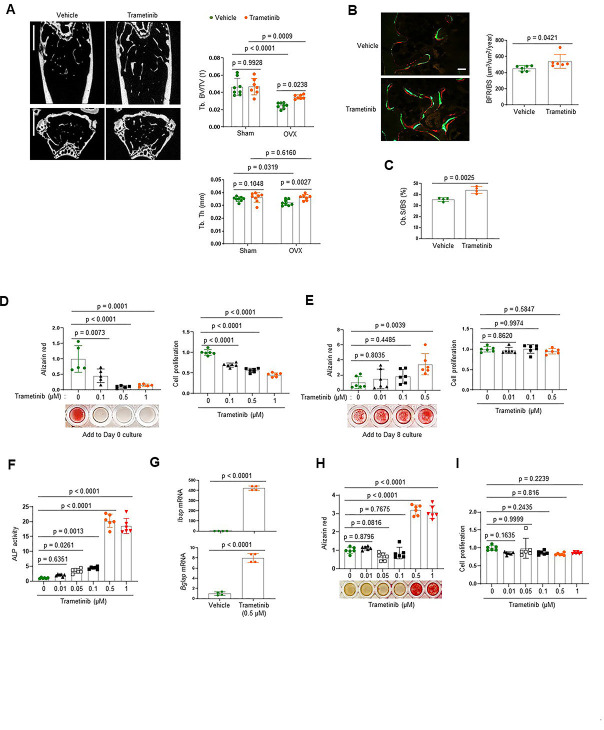
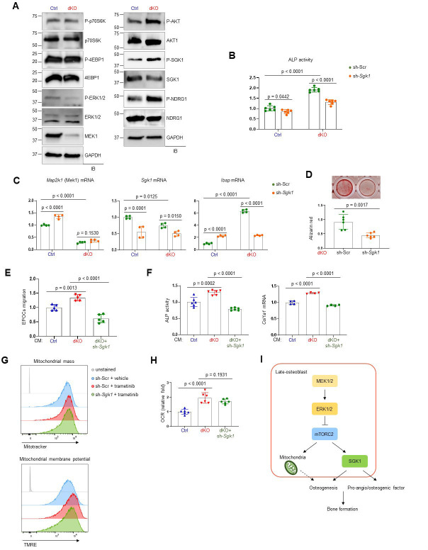
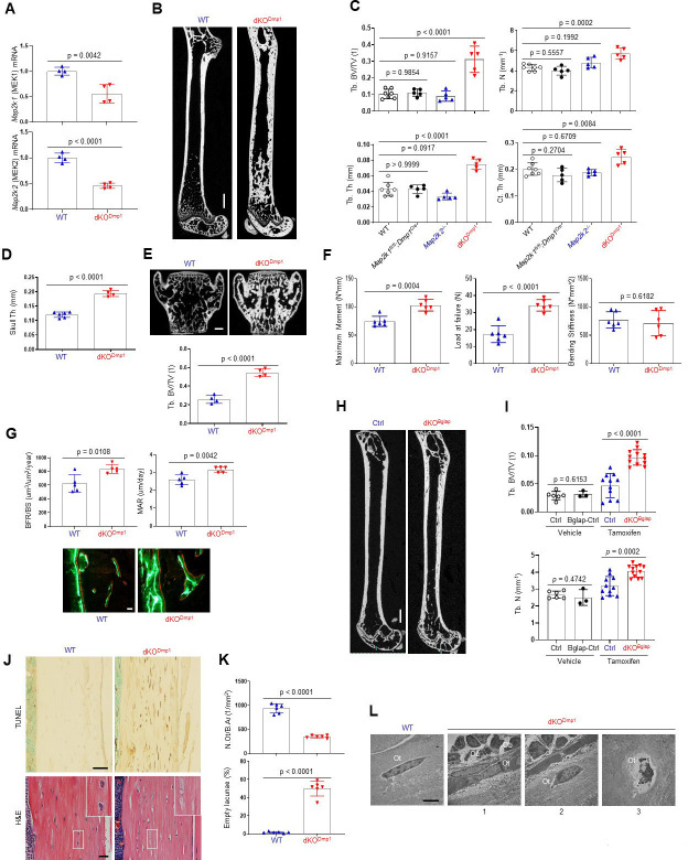
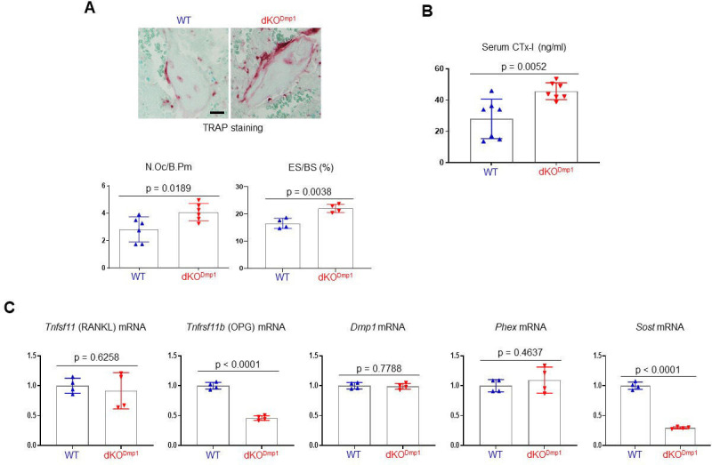
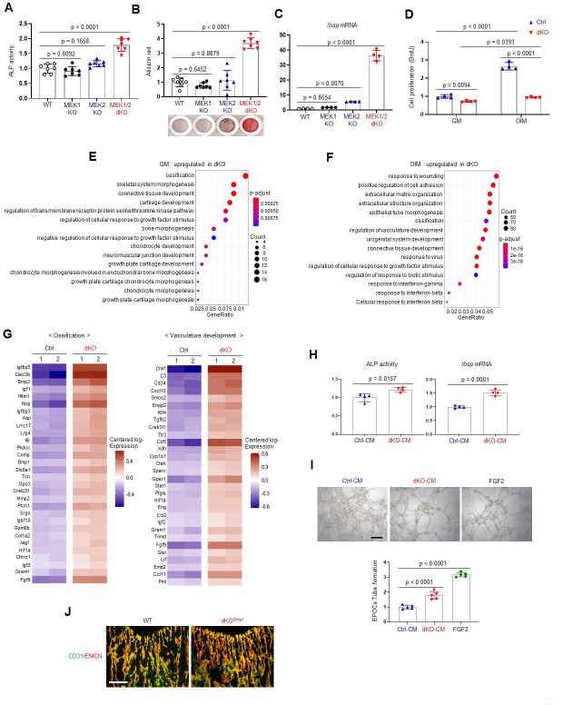
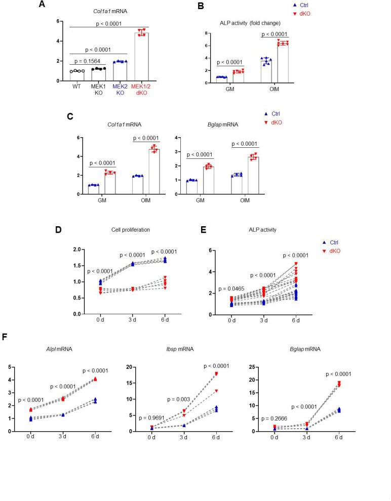
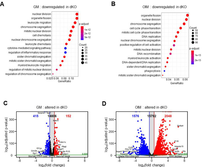
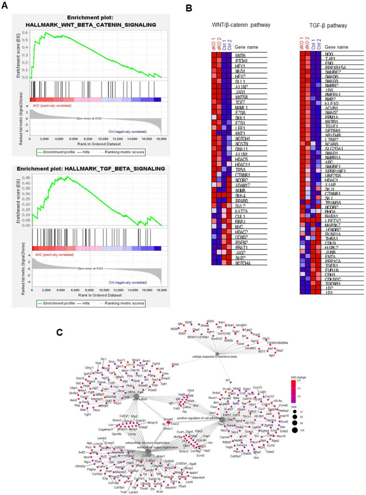
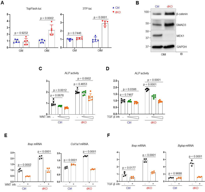
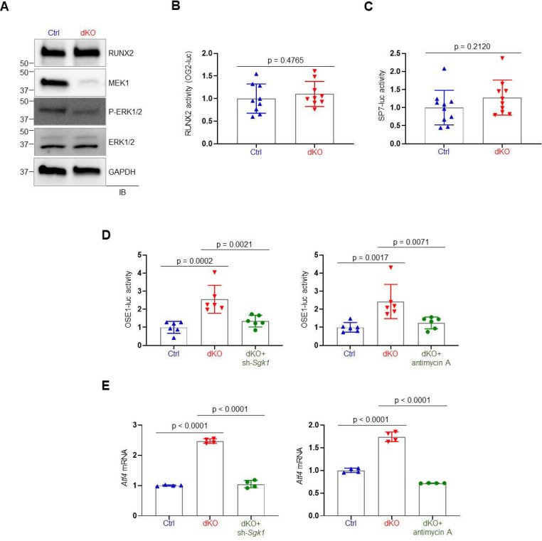
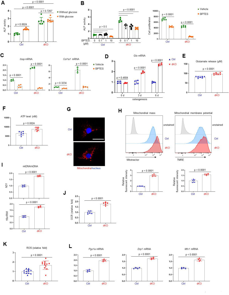
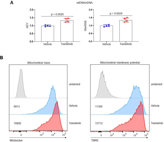
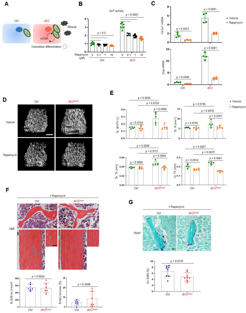
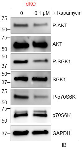
Emerging evidence supports that osteogenic differentiation of skeletal progenitors is a key determinant of overall bone formation and bone mass. Despite extensive studies showing the function of mitogen-activated protein kinases (MAPKs) in osteoblast differentiation, none of these studies show in vivo evidence of a role for MAPKs in osteoblast maturation subsequent to lineage commitment. Here, we describe how the extracellular signal-regulated kinase (ERK) pathway in osteoblasts controls bone formation by suppressing the mechanistic target of rapamycin (mTOR) pathway. We also show that, while ERK inhibition blocks the differentiation of osteogenic precursors when initiated at an early stage, ERK inhibition surprisingly promotes the later stages of osteoblast differentiation. Accordingly, inhibition of the ERK pathway using a small compound inhibitor or conditional deletion of the MAP2Ks (MEK1) and (MEK2), in mature osteoblasts and osteocytes, markedly increased bone formation due to augmented osteoblast differentiation. Mice with inducible deletion of the ERK pathway in mature osteoblasts also displayed similar phenotypes, demonstrating that this phenotype reflects continuous postnatal inhibition of late-stage osteoblast maturation. Mechanistically, ERK inhibition increases mitochondrial function and SGK1 phosphorylation via mTOR2 activation, which leads to osteoblast differentiation and production of angiogenic and osteogenic factors to promote bone formation. This phenotype was partially reversed by inhibiting mTOR. Our study uncovers a surprising dichotomy of ERK pathway functions in osteoblasts, whereby ERK activation promotes the early differentiation of osteoblast precursors, but inhibits the subsequent differentiation of committed osteoblasts via mTOR-mediated regulation of mitochondrial function and SGK1.

摘要

新出现的证据支持骨骼祖细胞的成骨分化是整体骨形成和骨量的关键决定因素。尽管有大量研究表明丝裂原活化蛋白激酶(MAPK)在成骨细胞分化中的作用,但这些研究都没有显示 MAPK 在谱系决定后对成骨细胞成熟的体内作用的证据。在这里,我们描述了成骨细胞中的细胞外信号调节激酶(ERK)途径如何通过抑制雷帕霉素靶蛋白(mTOR)途径来控制骨形成。我们还表明,虽然 ERK 抑制在早期阶段启动时会阻止成骨前体细胞的分化,但 ERK 抑制出人意料地促进了成骨细胞分化的后期阶段。因此,使用小化合物抑制剂或条件性删除 MAP2Ks(MEK1 和 MEK2)在成熟的成骨细胞和破骨细胞中抑制 ERK 途径,显著增加了骨形成,这是由于成骨细胞分化的增加。在成熟的成骨细胞中诱导缺失 ERK 途径的小鼠也表现出类似的表型,这表明这种表型反映了出生后晚期成骨细胞成熟的持续抑制。从机制上讲,ERK 抑制通过 mTOR2 激活增加线粒体功能和 SGK1 磷酸化,从而导致成骨细胞分化和产生血管生成和成骨因子以促进骨形成。这种表型部分通过抑制 mTOR 得到逆转。我们的研究揭示了 ERK 途径在成骨细胞中的作用的惊人二分法,即 ERK 激活促进成骨前体细胞的早期分化,但通过 mTOR 介导的对线粒体功能和 SGK1 的调节抑制后续分化。

https://cdn.ncbi.nlm.nih.gov/pmc/blobs/e6fe/9417416/acd1a9394f32/elife-78069-fig6.jpg
https://cdn.ncbi.nlm.nih.gov/pmc/blobs/e6fe/9417416/c3acd6b68276/elife-78069-fig1.jpg
https://cdn.ncbi.nlm.nih.gov/pmc/blobs/e6fe/9417416/1df5e072bf2f/elife-78069-fig1-figsupp1.jpg
https://cdn.ncbi.nlm.nih.gov/pmc/blobs/e6fe/9417416/f27daf7fdec5/elife-78069-fig2.jpg
https://cdn.ncbi.nlm.nih.gov/pmc/blobs/e6fe/9417416/f991835f6951/elife-78069-fig2-figsupp1.jpg
https://cdn.ncbi.nlm.nih.gov/pmc/blobs/e6fe/9417416/cb986e3f437b/elife-78069-fig3.jpg
https://cdn.ncbi.nlm.nih.gov/pmc/blobs/e6fe/9417416/f2b2f7f8d21a/elife-78069-fig3-figsupp1.jpg
https://cdn.ncbi.nlm.nih.gov/pmc/blobs/e6fe/9417416/18096103c8a3/elife-78069-fig3-figsupp2.jpg
https://cdn.ncbi.nlm.nih.gov/pmc/blobs/e6fe/9417416/fdbf39f487f4/elife-78069-fig3-figsupp3.jpg
https://cdn.ncbi.nlm.nih.gov/pmc/blobs/e6fe/9417416/17e6cacd50ab/elife-78069-fig3-figsupp4.jpg
https://cdn.ncbi.nlm.nih.gov/pmc/blobs/e6fe/9417416/006d350f9952/elife-78069-fig3-figsupp5.jpg
https://cdn.ncbi.nlm.nih.gov/pmc/blobs/e6fe/9417416/2f3659dc140c/elife-78069-fig4.jpg
https://cdn.ncbi.nlm.nih.gov/pmc/blobs/e6fe/9417416/e07c2d618300/elife-78069-fig4-figsupp1.jpg
https://cdn.ncbi.nlm.nih.gov/pmc/blobs/e6fe/9417416/ea1d1125a6bc/elife-78069-fig5.jpg
https://cdn.ncbi.nlm.nih.gov/pmc/blobs/e6fe/9417416/7ab7f8dc9e4c/elife-78069-fig5-figsupp1.jpg
https://cdn.ncbi.nlm.nih.gov/pmc/blobs/e6fe/9417416/acd1a9394f32/elife-78069-fig6.jpg

相似文献

1
Biphasic regulation of osteoblast development via the ERK MAPK-mTOR pathway.通过 ERK MAPK-mTOR 通路双向调节成骨细胞的发育。
Elife. 2022 Aug 17;11:e78069. doi: 10.7554/eLife.78069.
2
The ERK MAPK Pathway Is Essential for Skeletal Development and Homeostasis.ERK MAPK 通路对于骨骼发育和稳态至关重要。
Int J Mol Sci. 2019 Apr 12;20(8):1803. doi: 10.3390/ijms20081803.
3
TGF-β inhibits osteogenesis by upregulating the expression of ubiquitin ligase SMURF1 via MAPK-ERK signaling.转化生长因子-β通过丝裂原活化蛋白激酶-细胞外信号调节激酶信号通路上调泛素连接酶SMURF1的表达,从而抑制骨生成。
J Cell Physiol. 2018 Jan;233(1):596-606. doi: 10.1002/jcp.25920. Epub 2017 Jun 5.
4
EGFR controls bone development by negatively regulating mTOR-signaling during osteoblast differentiation.EGFR 通过负向调控成骨细胞分化过程中的 mTOR 信号来控制骨发育。
Cell Death Differ. 2018 Jun;25(6):1094-1106. doi: 10.1038/s41418-017-0054-7. Epub 2018 Feb 14.
5
Osteoblast-targeted suppression of PPARγ increases osteogenesis through activation of mTOR signaling.靶向成骨细胞抑制PPARγ通过激活mTOR信号通路增加骨生成。
Stem Cells. 2013 Oct;31(10):2183-92. doi: 10.1002/stem.1455.
6
MKP1-dependent PTH modulation of bone matrix mineralization in female mice is osteoblast maturation stage specific and involves P-ERK and P-p38 MAPKs.MKP1 依赖性甲状旁腺激素对雌性小鼠骨基质矿化的调节具有成骨细胞成熟阶段特异性,并涉及 P-ERK 和 P-p38 MAPKs。
J Endocrinol. 2013 Feb 25;216(3):315-29. doi: 10.1530/JOE-12-0372. Print 2013 Mar.
7
Mitogen-activated protein kinase pathways in osteoblasts.成骨细胞中的丝裂原活化蛋白激酶途径。
Annu Rev Cell Dev Biol. 2013;29:63-79. doi: 10.1146/annurev-cellbio-101512-122347. Epub 2013 May 31.
8
BMAL1 regulates osteoblast differentiation through mTOR/GSK3β/β-catenin pathway.BMAL1通过mTOR/GSK3β/β-连环蛋白通路调节成骨细胞分化。
J Mol Endocrinol. 2023 Apr 19;70(4). doi: 10.1530/JME-22-0181. Print 2023 May 1.
9
Estradiol inhibits osteoblast apoptosis via promotion of autophagy through the ER-ERK-mTOR pathway.雌二醇通过 ER-ERK-mTOR 通路促进自噬来抑制成骨细胞凋亡。
Apoptosis. 2013 Nov;18(11):1363-1375. doi: 10.1007/s10495-013-0867-x.
10
Oncostatin M receptor regulates osteoblast differentiation via extracellular signal-regulated kinase/autophagy signaling.抑瘤素 M 受体通过细胞外信号调节激酶/自噬信号通路调节成骨细胞分化。
Stem Cell Res Ther. 2022 Jun 28;13(1):278. doi: 10.1186/s13287-022-02958-1.

引用本文的文献

1
Reduced somatosensory innervation alters the skeletal transcriptome at a single cell level in a mouse model of type 2 diabetes.在2型糖尿病小鼠模型中,感觉神经支配减少会在单细胞水平上改变骨骼转录组。
Bone Res. 2025 Jul 4;13(1):67. doi: 10.1038/s41413-025-00436-x.
2
Activation of GLP-1 receptors enhances the osteogenic differentiation process of STRO-1-positive BMSCs.胰高血糖素样肽-1受体的激活增强了STRO-1阳性骨髓间充质干细胞的成骨分化过程。
Mol Biol Rep. 2025 Jun 6;52(1):559. doi: 10.1007/s11033-025-10648-3.
3
Unraveling the Potential of SGK1 in Osteoporosis: From Molecular Mechanisms to Therapeutic Targets.

本文引用的文献

1
The Extracellular Signal-Regulated Kinase Mitogen-Activated Protein Kinase Pathway in Osteoblasts.成骨细胞中的细胞外信号调节激酶丝裂原活化蛋白激酶信号通路
J Bone Metab. 2022 Feb;29(1):1-15. doi: 10.11005/jbm.2022.29.1.1. Epub 2022 Feb 28.
2
Vemurafenib plus Rituximab in Refractory or Relapsed Hairy-Cell Leukemia.维莫非尼联合利妥昔单抗治疗难治或复发的毛细胞白血病。
N Engl J Med. 2021 May 13;384(19):1810-1823. doi: 10.1056/NEJMoa2031298.
3
The Act of Controlling Adult Stem Cell Dynamics: Insights from Animal Models.控制成人干细胞动力学的行为:来自动物模型的见解。
揭示SGK1在骨质疏松症中的潜力:从分子机制到治疗靶点
Biomolecules. 2025 May 8;15(5):686. doi: 10.3390/biom15050686.
4
Nonessential amino acid is not nonessential in geriatric patients: implications for maxillofacial wound healing and bone repair.非必需氨基酸在老年患者中并非不重要:对颌面伤口愈合和骨修复的影响。
Maxillofac Plast Reconstr Surg. 2025 May 26;47(1):12. doi: 10.1186/s40902-025-00465-w.
5
Knockdown of IER3 Promotes Osteogenic Differentiation of Human Mesenchymal Stem Cells.IER3基因敲低促进人间充质干细胞的成骨分化。
Biomedicines. 2025 Apr 12;13(4):947. doi: 10.3390/biomedicines13040947.
6
[4‑(Arylethyl)‑pyrrolo[2,3-d] pyrimidine improves post-traumatic stress disorder in mice by inhibiting mGluR5-regulated ERK1/2-SGK1 signaling pathway].[4-(芳基乙基)-吡咯并[2,3-d]嘧啶通过抑制代谢型谷氨酸受体5调节的细胞外信号调节激酶1/2-血清/糖皮质激素调节激酶1信号通路改善小鼠创伤后应激障碍]
Nan Fang Yi Ke Da Xue Xue Bao. 2025 Apr 20;45(4):765-773. doi: 10.12122/j.issn.1673-4254.2025.04.12.
7
Molecular Evidence Supporting MEK Inhibitor Therapy in NF1 Pseudarthrosis.支持MEK抑制剂治疗神经纤维瘤病1型假关节的分子证据。
J Bone Joint Surg Am. 2025 May 21;107(10):1098-1106. doi: 10.2106/JBJS.24.01007. Epub 2025 Apr 1.
8
MK-4 Ameliorates Diabetic Osteoporosis in Angiogenesis-Dependent Bone Formation by Promoting Mitophagy in Endothelial Cells.MK-4通过促进内皮细胞中的线粒体自噬改善血管生成依赖性骨形成中的糖尿病性骨质疏松症。
Drug Des Devel Ther. 2025 Mar 25;19:2173-2188. doi: 10.2147/DDDT.S503930. eCollection 2025.
9
Investigating the Role of Shikonin in Enhancing Osteogenesis and Angiogenesis for the Treatment of Osteoporosis.研究紫草素在增强成骨作用和血管生成以治疗骨质疏松症中的作用。
ACS Omega. 2025 Feb 27;10(9):9718-9727. doi: 10.1021/acsomega.4c11161. eCollection 2025 Mar 11.
10
Impaired MC3T3-E1 osteoblast differentiation triggered by oncogenic HRAS is rescued by the farnesyltransferase inhibitor Tipifarnib.致癌性HRAS引发的MC3T3-E1成骨细胞分化受损可被法尼基转移酶抑制剂替匹法尼挽救。
Sci Rep. 2025 Feb 26;15(1):6832. doi: 10.1038/s41598-025-91592-x.
Biomolecules. 2021 Apr 30;11(5):667. doi: 10.3390/biom11050667.
4
The bone microenvironment increases phenotypic plasticity of ER breast cancer cells.骨微环境增加了雌激素受体阳性乳腺癌细胞的表型可塑性。
Dev Cell. 2021 Apr 19;56(8):1100-1117.e9. doi: 10.1016/j.devcel.2021.03.008.
5
The bone microenvironment invigorates metastatic seeds for further dissemination.骨微环境激活转移种子以促进进一步扩散。
Cell. 2021 Apr 29;184(9):2471-2486.e20. doi: 10.1016/j.cell.2021.03.011. Epub 2021 Apr 19.
6
SGK1 signaling promotes glucose metabolism and survival in extracellular matrix detached cells.SGK1信号传导促进细胞外基质脱离细胞中的葡萄糖代谢和存活。
Cell Rep. 2021 Mar 16;34(11):108821. doi: 10.1016/j.celrep.2021.108821.
7
Regulation and metabolic functions of mTORC1 and mTORC2.mTORC1 和 mTORC2 的调节和代谢功能。
Physiol Rev. 2021 Jul 1;101(3):1371-1426. doi: 10.1152/physrev.00026.2020. Epub 2021 Feb 18.
8
Rictor/mTORC2 involves mitochondrial function in ES cells derived cardiomyocytes via mitochondrial Connexin 43.雷帕霉素靶蛋白复合体 2(Rictor/mTORC2)通过线粒体间隙连接蛋白 43(Connexin 43)参与胚胎干细胞衍生心肌细胞的线粒体功能。
Acta Pharmacol Sin. 2021 Nov;42(11):1790-1797. doi: 10.1038/s41401-020-00591-3. Epub 2021 Feb 5.
9
Genetic mechanisms of critical illness in COVID-19.新型冠状病毒肺炎危重症的遗传机制。
Nature. 2021 Mar;591(7848):92-98. doi: 10.1038/s41586-020-03065-y. Epub 2020 Dec 11.
10
Overcoming Adaptive Resistance to KRAS and MEK Inhibitors by Co-targeting mTORC1/2 Complexes in Pancreatic Cancer.通过共同靶向胰腺癌中的 mTORC1/2 复合物克服 KRAS 和 MEK 抑制剂的适应性耐药性。
Cell Rep Med. 2020 Nov 17;1(8):100131. doi: 10.1016/j.xcrm.2020.100131.