Institute of Dentistry, University of Eastern Finland, Yliopistonranta 1, Kuopio, 70210, Finland.
SIB Labs, University of Eastern Finland, Yliopistonranta 1, Kuopio, 70210, Finland.
Calcif Tissue Int. 2022 Dec;111(6):547-558. doi: 10.1007/s00223-022-01017-4. Epub 2022 Aug 17.
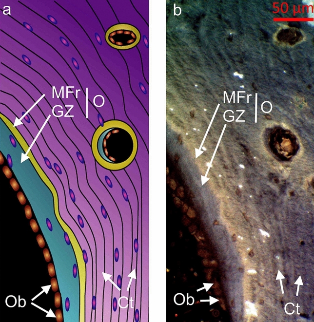
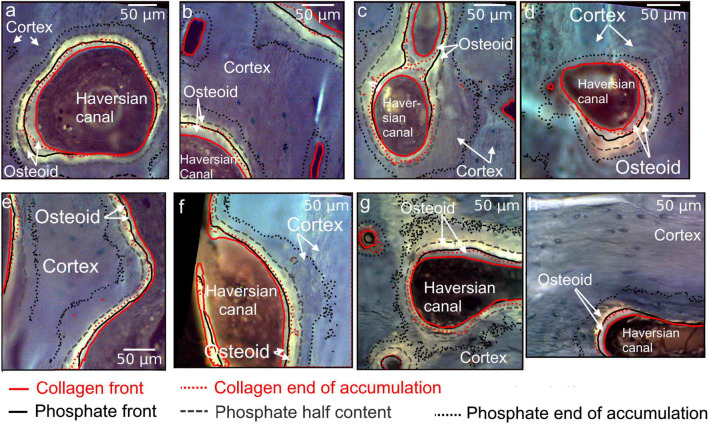
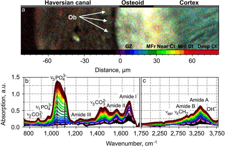
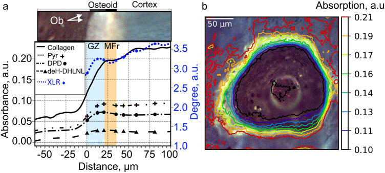
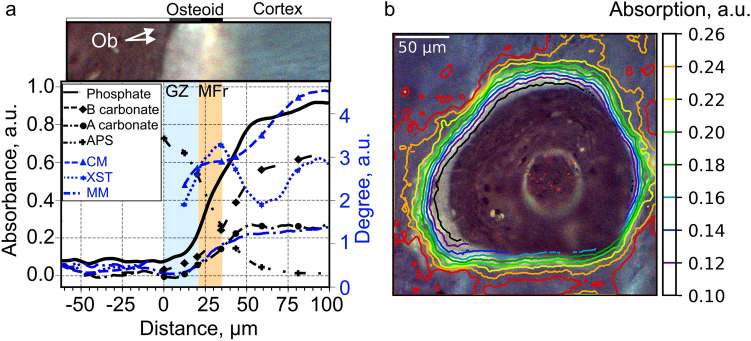
Osteoid is a layer of new-formed bone that is deposited on the bone border during the process of new bone formation. This deposition process is crucial for bone tissue, and flaws in it can lead to bone diseases. Certain bone diseases, i.e. medication related osteonecrosis, are overexpressed in mandibular bone. Because mandibular bone presents different properties than other bone types, the data concerning osteoid formation in other bones are inapplicable for human-mandibular bone. Previously, the molecular distribution of other bone types has been presented using Fourier-transform infrared (FTIR) spectroscopy. However, the spatial distribution of molecular components of healthy-human-mandibular-bone osteoid in relation to histologic landmarks has not been previously presented and needs to be studied in order to understand diseases that occur human-mandibular bone. This study presents for the first time the variation in molecular distribution inside healthy-human-mandibular-bone osteoid by juxtaposing FTIR data with its corresponding histologic image obtained by autofluorescence imaging of its same bone section. During new bone formation, bone-forming cells produce an osteoid constituted primarily of type I collagen. It was observed that in mandibular bone, the collagen type I increases from the osteoblast line with the distance from the osteoblasts, indicating progressive accumulation of collagen during osteoid formation. Only later inside the collagen matrix, the osteoid starts to mineralize. When the mineralization starts, the collagen accumulation diminishes whereas the collagen maturation still continues. This chemical-apposition process in healthy mandibular bone will be used in future as a reference to understand different pathologic conditions that occur in human-mandibular bone.
类骨质是在新骨形成过程中沉积在骨边界上的新形成的骨层。这种沉积过程对骨组织至关重要,其缺陷会导致骨病。某些骨病,如与药物相关的骨坏死,在下颌骨中过度表达。由于下颌骨与其他类型的骨骼具有不同的特性,因此其他骨骼中类骨质形成的数据不适用于人类下颌骨。以前,已经使用傅里叶变换红外(FTIR)光谱法呈现了其他类型骨骼的分子分布。然而,健康人类下颌骨类骨质的分子成分的空间分布与组织学标志之间的关系以前尚未呈现出来,需要进行研究,以便了解发生在人类下颌骨中的疾病。本研究首次通过将 FTIR 数据与其同一骨切片的自发荧光成像获得的相应组织学图像并列,呈现了健康人类下颌骨类骨质内部分子分布的变化。在新骨形成过程中,成骨细胞产生主要由 I 型胶原组成的类骨质。观察到在下颌骨中,胶原 I 型从成骨细胞线随距离成骨细胞的增加而增加,表明在类骨质形成过程中胶原逐渐积累。只有在胶原基质内部,类骨质才开始矿化。当矿化开始时,胶原的积累减少,而胶原的成熟仍在继续。这种健康下颌骨中的化学附着过程将在未来作为理解发生在人类下颌骨中的不同病理状况的参考。