• 文献检索
  • 文档翻译
  • 深度研究
  • 学术资讯
  • Suppr Zotero 插件Zotero 插件
  • 邀请有礼
  • 套餐&价格
  • 历史记录
应用&插件
Suppr Zotero 插件Zotero 插件浏览器插件Mac 客户端Windows 客户端微信小程序
定价
高级版会员购买积分包购买API积分包
服务
文献检索文档翻译深度研究API 文档MCP 服务
关于我们
关于 Suppr公司介绍联系我们用户协议隐私条款
关注我们

Suppr 超能文献

核心技术专利:CN118964589B侵权必究
粤ICP备2023148730 号-1Suppr @ 2026

文献检索

告别复杂PubMed语法,用中文像聊天一样搜索,搜遍4000万医学文献。AI智能推荐,让科研检索更轻松。

立即免费搜索

文件翻译

保留排版,准确专业,支持PDF/Word/PPT等文件格式,支持 12+语言互译。

免费翻译文档

深度研究

AI帮你快速写综述,25分钟生成高质量综述,智能提取关键信息,辅助科研写作。

立即免费体验

Semaphorin3f 作为一种心肌细胞来源的心脏室发育调节剂。

Semaphorin3f as a cardiomyocyte derived regulator of heart chamber development.

机构信息

Hotchkiss Brain Institute, Alberta Children's Hospital Research Institute, Department of Cell Biology and Anatomy, University of Calgary, 3330 Hospital Dr., NW, Calgary, AB, T2N 4N1, Canada.

出版信息

Cell Commun Signal. 2022 Aug 19;20(1):126. doi: 10.1186/s12964-022-00874-8.

DOI:10.1186/s12964-022-00874-8
PMID:35986301
原文链接:https://pmc.ncbi.nlm.nih.gov/articles/PMC9389736/
Abstract

BACKGROUND

During development a pool of precursors form a heart with atrial and ventricular chambers that exhibit distinct transcriptional and electrophysiological properties. Normal development of these chambers is essential for full term survival of the fetus, and deviations result in congenital heart defects. The large number of genes that may cause congenital heart defects when mutated, and the genetic variability and penetrance of the ensuing phenotypes, reveals a need to understand the molecular mechanisms that allow for the formation of chamber-specific cardiomyocyte differentiation.

METHODS

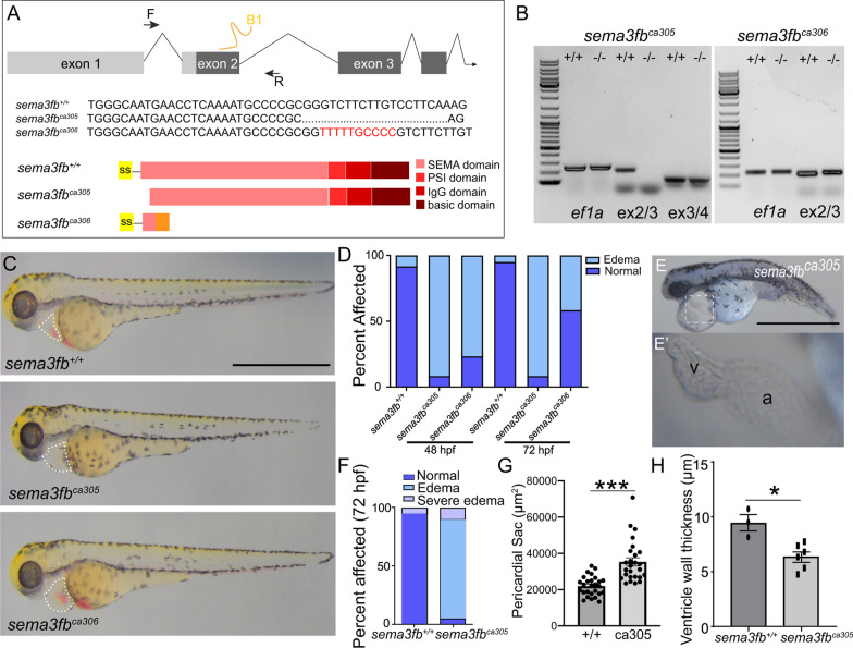
We used in situ hybridization, immunohistochemistry and functional analyses to identify the consequences of the loss of the secreted semaphorin, Sema3fb, in the development of the zebrafish heart by using two sema3fb CRISPR mutant alleles.

RESULTS

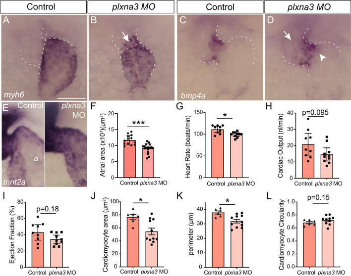
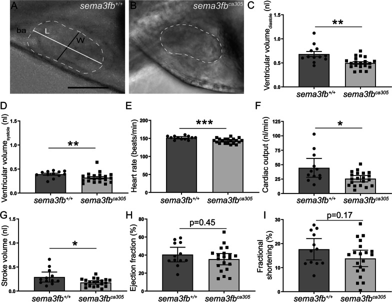
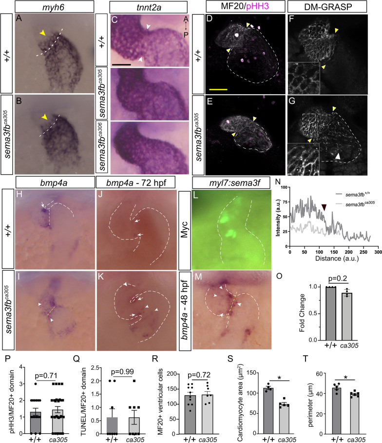
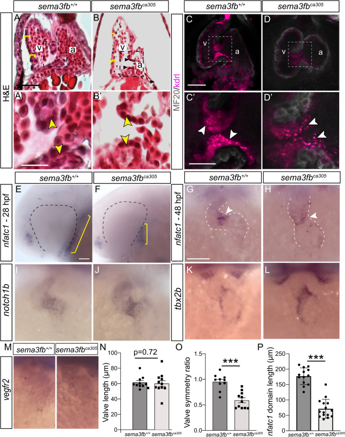
We find that in the developing zebrafish heart sema3fb mRNA is expressed by all cardiomyocytes, whereas mRNA for a known receptor Plexina3 (Plxna3) is expressed preferentially by ventricular cardiomyocytes. In sema3fb CRISPR zebrafish mutants, heart chamber development is impaired; the atria and ventricles of mutants are smaller in size than their wild type siblings, apparently because of differences in cell size and not cell numbers. Analysis of chamber differentiation indicates defects in chamber specific gene expression at the border between the ventricular and atrial chambers, with spillage of ventricular chamber genes into the atrium, and vice versa, and a failure to restrict specialized cardiomyocyte markers to the atrioventricular canal (AVC). The hypoplastic heart chambers are associated with decreased cardiac output and heart edema.

CONCLUSIONS

Based on our data we propose a model whereby cardiomyocytes secrete a Sema cue that, because of spatially restricted expression of the receptor, signals in a ventricular chamber-specific manner to establish a distinct border between atrial and ventricular chambers that is important to produce a fully functional heart. Video abstract.

摘要

背景

在发育过程中,一群前体细胞形成具有明显转录和电生理特性的心房和心室腔的心脏。这些腔室的正常发育对于胎儿的足月存活至关重要,而发育偏差则导致先天性心脏缺陷。大量可能导致先天性心脏缺陷的基因突变,以及随之而来的表型的遗传变异性和外显率,揭示了需要了解允许形成腔特异性心肌细胞分化的分子机制。

方法

我们使用原位杂交、免疫组织化学和功能分析来鉴定两个 sema3fb CRISPR 突变等位基因在斑马鱼心脏发育过程中缺失分泌的信号素 Sema3fb 的后果。

结果

我们发现,在发育中的斑马鱼心脏中,sema3fb mRNA 由所有心肌细胞表达,而已知受体 Plexina3(Plxna3)的 mRNA 则优先由心室心肌细胞表达。在 sema3fb CRISPR 斑马鱼突变体中,心脏腔室发育受损;突变体的心房和心室比其野生型兄弟姐妹小,显然是由于细胞大小的差异而不是细胞数量的差异。对腔室分化的分析表明,在心室和心房腔之间的边界处存在腔特异性基因表达缺陷,心室腔基因溢出到心房,反之亦然,并且不能将专门的心肌细胞标记物限制在房室管(AVC)内。发育不良的心脏腔室与心输出量减少和心脏水肿有关。

结论

基于我们的数据,我们提出了一个模型,即心肌细胞分泌一种信号素 cue,由于受体的空间限制表达,以心室腔特异性的方式发出信号,在心房和心室腔之间建立一个重要的独特边界,这对于产生一个功能齐全的心脏至关重要。视频摘要。

https://cdn.ncbi.nlm.nih.gov/pmc/blobs/c61b/9389736/c9a1861b363a/12964_2022_874_Fig7_HTML.jpg
https://cdn.ncbi.nlm.nih.gov/pmc/blobs/c61b/9389736/fd2530d204de/12964_2022_874_Fig1_HTML.jpg
https://cdn.ncbi.nlm.nih.gov/pmc/blobs/c61b/9389736/c7bca5b633bb/12964_2022_874_Fig2_HTML.jpg
https://cdn.ncbi.nlm.nih.gov/pmc/blobs/c61b/9389736/db586b4d12fa/12964_2022_874_Fig3_HTML.jpg
https://cdn.ncbi.nlm.nih.gov/pmc/blobs/c61b/9389736/4af7822faf4a/12964_2022_874_Fig4_HTML.jpg
https://cdn.ncbi.nlm.nih.gov/pmc/blobs/c61b/9389736/d34765d1b87c/12964_2022_874_Fig5_HTML.jpg
https://cdn.ncbi.nlm.nih.gov/pmc/blobs/c61b/9389736/8d4ae69584e2/12964_2022_874_Fig6_HTML.jpg
https://cdn.ncbi.nlm.nih.gov/pmc/blobs/c61b/9389736/c9a1861b363a/12964_2022_874_Fig7_HTML.jpg

相似文献

1
Semaphorin3f as a cardiomyocyte derived regulator of heart chamber development.Semaphorin3f 作为一种心肌细胞来源的心脏室发育调节剂。
Cell Commun Signal. 2022 Aug 19;20(1):126. doi: 10.1186/s12964-022-00874-8.
2
Nkx genes are essential for maintenance of ventricular identity.Nkx 基因对于维持心室身份至关重要。
Development. 2013 Oct;140(20):4203-13. doi: 10.1242/dev.095562. Epub 2013 Sep 11.
3
Nr2f1a balances atrial chamber and atrioventricular canal size via BMP signaling-independent and -dependent mechanisms.Nr2f1a通过不依赖和依赖骨形态发生蛋白(BMP)信号传导的机制来平衡心房腔和房室管的大小。
Dev Biol. 2018 Feb 1;434(1):7-14. doi: 10.1016/j.ydbio.2017.11.010. Epub 2017 Nov 20.
4
Gon4l/Udu regulates cardiomyocyte proliferation and maintenance of ventricular chamber identity during zebrafish development.Gon4l/Udu 在斑马鱼发育过程中调节心肌细胞增殖和心室腔身份的维持。
Dev Biol. 2020 Jun 15;462(2):223-234. doi: 10.1016/j.ydbio.2020.03.002. Epub 2020 Apr 6.
5
Chamber Specific Gene Expression Landscape of the Zebrafish Heart.斑马鱼心脏的腔室特异性基因表达图谱
PLoS One. 2016 Jan 27;11(1):e0147823. doi: 10.1371/journal.pone.0147823. eCollection 2016.
6
FGF signaling enforces cardiac chamber identity in the developing ventricle.成纤维细胞生长因子(FGF)信号传导在发育中的心室中维持心腔特征。
Development. 2017 Apr 1;144(7):1328-1338. doi: 10.1242/dev.143719. Epub 2017 Feb 23.
7
Mutation of weak atrium/atrial myosin heavy chain disrupts atrial function and influences ventricular morphogenesis in zebrafish.弱心房/心房肌球蛋白重链的突变会破坏斑马鱼的心房功能并影响心室形态发生。
Development. 2003 Dec;130(24):6121-9. doi: 10.1242/dev.00838. Epub 2003 Oct 22.
8
Alternative splicing of jnk1a in zebrafish determines first heart field ventricular cardiomyocyte numbers through modulation of hand2 expression.在斑马鱼中,jnk1a 的可变剪接通过调节 hand2 表达来决定第一心区心室心肌细胞的数量。
PLoS Genet. 2020 May 18;16(5):e1008782. doi: 10.1371/journal.pgen.1008782. eCollection 2020 May.
9
Heart chamber size in zebrafish is regulated redundantly by duplicated tbx2 genes.斑马鱼心脏腔室大小由重复的 tbx2 基因冗余调控。
Dev Dyn. 2011 Jun;240(6):1548-57. doi: 10.1002/dvdy.22622. Epub 2011 Mar 29.
10
Nr2f1a maintains atrial expression to repress pacemaker identity within venous atrial cardiomyocytes of zebrafish.Nr2f1a 通过维持心房表达来抑制斑马鱼静脉心房心肌细胞中的起搏细胞特征。
Elife. 2023 May 15;12:e77408. doi: 10.7554/eLife.77408.

引用本文的文献

1
Regionalized regulation of actomyosin organization influences cardiomyocyte cell shape changes during chamber curvature formation.肌动球蛋白组织的区域化调控影响心室曲率形成过程中心肌细胞的形态变化。
bioRxiv. 2025 Jan 8:2025.01.07.631779. doi: 10.1101/2025.01.07.631779.
2
Zebrafish Congenital Heart Disease Models: Opportunities and Challenges.斑马鱼先天性心脏病模型:机遇与挑战。
Int J Mol Sci. 2024 May 29;25(11):5943. doi: 10.3390/ijms25115943.

本文引用的文献

1
Zebrafish Models in Therapeutic Research of Cardiac Conduction Disease.斑马鱼模型在心脏传导疾病治疗研究中的应用
Front Cell Dev Biol. 2021 Aug 4;9:731402. doi: 10.3389/fcell.2021.731402. eCollection 2021.
2
Unveiling Complexity and Multipotentiality of Early Heart Fields.揭示早期心脏场的复杂性和多潜能性。
Circ Res. 2021 Aug 6;129(4):474-487. doi: 10.1161/CIRCRESAHA.121.318943. Epub 2021 Jun 24.
3
Semaphorin 3fa Controls Ocular Vascularization From the Embryo Through to the Adult.Semaphorin 3fa 调控胚胎到成年期的眼部血管生成。
Invest Ophthalmol Vis Sci. 2021 Feb 1;62(2):21. doi: 10.1167/iovs.62.2.21.
4
Llgl1 regulates zebrafish cardiac development by mediating Yap stability in cardiomyocytes.Llgl1 通过调节心肌细胞中 Yap 的稳定性来调控斑马鱼心脏发育。
Development. 2020 Aug 25;147(16):dev193581. doi: 10.1242/dev.193581.
5
Semaphorin 3F signaling actively retains neutrophils at sites of inflammation.信号蛋白 Semaphorin 3F 通过信号通路积极地将中性粒细胞保留在炎症部位。
J Clin Invest. 2020 Jun 1;130(6):3221-3237. doi: 10.1172/JCI130834.
6
Functional Heterogeneity within the Developing Zebrafish Epicardium.斑马鱼心脏外胚层的功能异质性。
Dev Cell. 2020 Mar 9;52(5):574-590.e6. doi: 10.1016/j.devcel.2020.01.023. Epub 2020 Feb 20.
7
Nfatc1 Promotes Interstitial Cell Formation During Cardiac Valve Development in Zebrafish.Nfatc1 促进斑马鱼心脏瓣膜发育过程中的间质细胞形成。
Circ Res. 2020 Apr 10;126(8):968-984. doi: 10.1161/CIRCRESAHA.119.315992. Epub 2020 Feb 18.
8
Vezf1 regulates cardiac structure and contractile function.VEZF1 调节心脏结构和收缩功能。
EBioMedicine. 2020 Jan;51:102608. doi: 10.1016/j.ebiom.2019.102608. Epub 2020 Jan 3.
9
Muscarinic receptors promote pacemaker fate at the expense of secondary conduction system tissue in zebrafish.毒蕈碱型乙酰胆碱受体通过牺牲次级传导系统组织促进斑马鱼起搏细胞的命运。
JCI Insight. 2019 Oct 17;4(20):121971. doi: 10.1172/jci.insight.121971.
10
Semaphorin 3E/PlexinD1 signaling is required for cardiac ventricular compaction.Semaphorin 3E/PlexinD1 信号通路对于心室致密化是必需的。
JCI Insight. 2019 Aug 22;4(16):125908. doi: 10.1172/jci.insight.125908.