Respiratory Translational Research Group, Department of Laboratory Medicine, School of Health Sciences, College of Health and Medicine, University of Tasmania, Launceston, Tasmania, Australia.
Department of Respiratory Medicine, Tasmanian Health Services (THS), North-West Hospital, Burnie, Tasmania, Australia.
Am J Physiol Lung Cell Mol Physiol. 2022 Oct 1;323(4):L473-L483. doi: 10.1152/ajplung.00137.2022. Epub 2022 Aug 23.
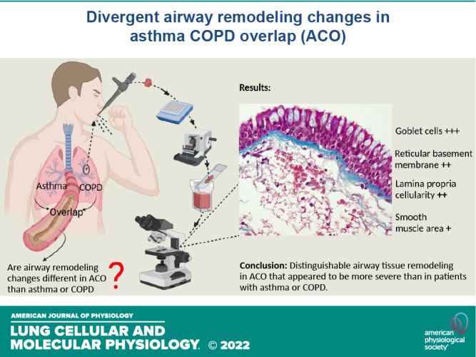
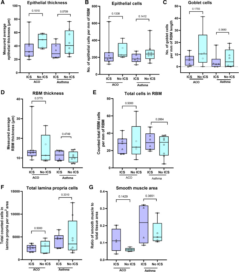
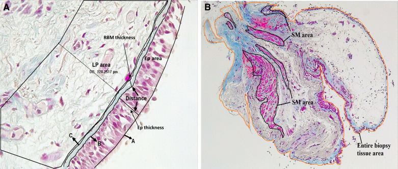
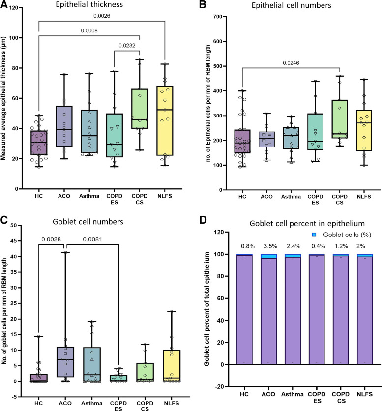
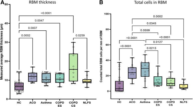
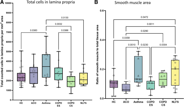
Management of patients with asthma COPD overlap (ACO) is clinically challenging due to insufficient evidence of pathological changes in these patients. In this cross-sectional study, we evaluated airway remodeling in endobronchial biopsies from a total of 90 subjects, which included 12 ACO, 14 patients with asthma, 12 COPD exsmokers (ES), 11 current smokers (CS), 28 healthy controls (HC), and 13 normal lung function smokers (NLFS). Tissue was stained with Masson's trichrome. Epithelium, goblet cells, reticular basement membrane (RBM), cellularity, lamina propria (LP), and smooth muscle (SM) changes were measured using Image-Pro Plus v7 software. Differential airway remodeling pattern was seen in patients with ACO. A limited change was noted in the ACO epithelium compared with other pathological groups. RBM was substantially thicker in patients with ACO than in HC ( < 0.0002) and tended to be thicker than in patients with asthma and NLFS. The total RBM cells were higher in ACO than in the HC ( < 0.0001), COPD-CS ( = 0.0559), -ES ( = 0.0345), and NLFS ( < 0.0002), but did not differ from patients with asthma. Goblet cells were higher in the ACO than in the HC ( = 0.0028) and COPD-ES ( = 0.0081). The total LP cells in ACO appeared to be higher than in HC, COPD-CS, and NLFS but appeared to be lower than in patients with asthma. Finally, SM area was significantly lower in the ACO than in patients with asthma ( = 0.001), COPD-CS (=0.0290), and NLFS ( = 0.0011). This first comprehensive study suggests that patients with ACO had distinguishable tissue remodeling that appeared to be more severe than patients with asthma and COPD. This study will help in informed decision-making for better patient management in clinical practice.
哮喘-慢性阻塞性肺疾病重叠(ACO)患者的管理具有临床挑战性,因为这些患者的病理变化证据不足。在这项横断面研究中,我们评估了总共 90 名受试者的支气管内膜活检中的气道重塑,其中包括 12 名 ACO 患者、14 名哮喘患者、12 名 COPD 戒烟者(ES)、11 名当前吸烟者(CS)、28 名健康对照者(HC)和 13 名正常肺功能吸烟者(NLFS)。组织用 Masson 三色染色。使用 Image-Pro Plus v7 软件测量上皮、杯状细胞、网状基底膜(RBM)、细胞数、固有层(LP)和平滑肌(SM)的变化。ACO 患者表现出不同的气道重塑模式。与其他病理组相比,ACO 患者的上皮变化有限。与 HC(<0.0002)相比,ACO 患者的 RBM 明显更厚,并倾向于比哮喘和 NLFS 患者的 RBM 更厚。与 HC(<0.0001)、COPD-CS(=0.0559)、-ES(=0.0345)和 NLFS(<0.0002)相比,ACO 患者的总 RBM 细胞更高,但与哮喘患者无差异。与 HC(=0.0028)和 COPD-ES(=0.0081)相比,ACO 患者的杯状细胞更高。ACO 患者的总 LP 细胞似乎高于 HC、COPD-CS 和 NLFS,但似乎低于哮喘患者。最后,与哮喘患者(=0.001)、COPD-CS(=0.0290)和 NLFS(=0.0011)相比,ACO 患者的 SM 面积明显更低。这项首次全面研究表明,ACO 患者的组织重塑具有可识别的特征,似乎比哮喘和 COPD 患者更严重。这项研究将有助于在临床实践中为更好的患者管理做出明智的决策。