Department of Surgery, Dentistry, Gynecology and Pediatrics, Division of General and Hepato-Biliary Surgery, University of Verona, P. le L.A. Scuro 10, 37134, Verona, Italy.
Updates Surg. 2023 Jan;75(1):105-114. doi: 10.1007/s13304-022-01365-8. Epub 2022 Aug 25.
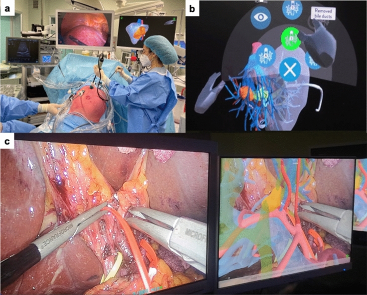
Three-dimensional visualization technology (3DVT) has been recently introduced to achieve a precise preoperative planning of liver surgery. The aim of this observational study was to assess the accuracy of 3DVT for complex liver resections. 3DVT with hyper accuracy three-dimensional (HA3D™) technology was introduced at our institution on February 2020. Anatomical characteristics were collected from two-dimensional imaging (2DI) and 3DVT, while intraoperative and postoperative outcomes were recorded prospectively. A total of 62 patients were enrolled into the study. 3DVT was able to study tumor extension and liver anatomy, identifying at least one vascular variation in 37 patients (59.7%). Future remnant liver volume (FRLV) was measured using 2DI and 3DVT. The paired samples t test assessed positive correlation between the two methods (p < 0.001). At least one vessel was suspected to be invaded by the tumor in 8 (15.7%) 2DI cases vs 16 (31.4%) 3DVT cases, respectively. During surgery, vascular invasion was detected in 17 patients (33.3%). A total of 73 surgical procedures were proposed basing on 2DI, including 2 alternatives for 16 patients. After 3DVT, the previously planned procedure was changed in 15 cases (29.4%), due to the clearer information provided. A total of 51 patients (82%) underwent surgery. The most frequent procedure was right hepatectomy (33.3%), followed by left hepatectomy (23.5%) and left trisectionectomy (13.7%). Vascular resection and reconstruction were performed in 10 patients (19.6%) and portal vein was resected in more than half of these cases (66.7%). 3DVT leads to a more detailed and tailored approach to complex liver surgery, improving surgeons' knowledge of liver anatomy and accuracy of liver resection.
三维可视化技术(3DVT)最近被引入以实现肝切除术的精确术前规划。本观察性研究的目的是评估 3DVT 对复杂肝切除术的准确性。我们机构于 2020 年 2 月引入了具有超精确三维(HA3D™)技术的 3DVT。从二维成像(2DI)和 3DVT 中收集解剖特征,同时前瞻性记录术中及术后结果。共有 62 名患者入组本研究。3DVT 能够研究肿瘤的延伸和肝脏解剖,在 37 名患者(59.7%)中确定至少 1 种血管变异。使用 2DI 和 3DVT 测量未来剩余肝脏体积(FRLV)。配对样本 t 检验评估了两种方法之间的正相关性(p<0.001)。在 2DI 病例中,至少有 1 个血管被怀疑被肿瘤侵犯的有 8 例(15.7%),而在 3DVT 病例中则有 16 例(31.4%)。在手术中,17 名患者(33.3%)检测到血管侵犯。共有 73 例手术方案基于 2DI 提出,其中 16 名患者有 2 种替代方案。在进行 3DVT 后,由于提供了更清晰的信息,15 例(29.4%)之前计划的手术方案发生了改变。共有 51 名患者(82%)接受了手术。最常见的手术是右半肝切除术(33.3%),其次是左半肝切除术(23.5%)和左三叶切除术(13.7%)。10 名患者(19.6%)进行了血管切除和重建,其中超过一半的病例(66.7%)切除了门静脉。3DVT 可使复杂肝切除术更详细和更具针对性,提高外科医生对肝脏解剖的了解和肝切除术的准确性。