Department of Trauma and Orthopedic Surgery, Friedrich-Alexander-Universität Erlangen-Nürnberg, University Hospital Erlangen, Krankenhaus-street. 12, 91054, Erlangen, Germany.
BG Trauma Center Ludwigshafen at Ruprecht-Karls-Universität Heidelberg, Ludwig-Guttmann-Street. 13, 67071, Ludwigshafen, Germany.
Eur J Trauma Emerg Surg. 2023 Feb;49(1):373-381. doi: 10.1007/s00068-022-02083-x. Epub 2022 Sep 1.
Intraoperative 3D imaging has become a valued tool in assessing the quality of reduction and implant placement in orthopedic trauma surgery. In our institution, 3D imaging is used routinely since 2001. To evaluate the intraoperative findings and consequences of this technique, intraoperative revision rates in cases with 3D imaging were analyzed.
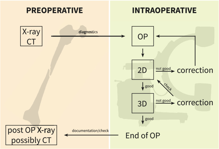
All operative procedures carried out with intraoperative 3D imaging between August 2001 and December 2016 were included. The scans were assessed intraoperatively and documented thereafter. In case of malreduction or misplaced implants, an immediate revision was performed. The number of scans per case as well as the findings and consequences drawn regarding the anatomical region were analyzed.
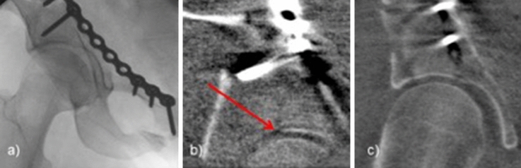
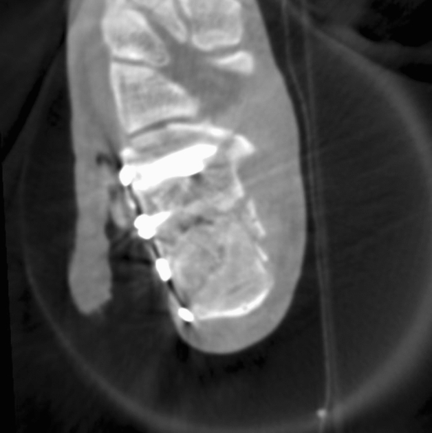
4721 cases with 7201 3D scans were included in this study. The most common anatomical regions were the ankle (22.3%), the calcaneus (14.8%) and the tibial head (9.5%). In 19.1% of all cases, an intraoperative revision was performed. The highest revision rates were found with 36.0% in calcaneal fractures, 24.8% in fractures of the tibial plateau, 22.3% in injuries of the ankle. In 52.0% of revisions, the reduction was improved regarding intra-articular steps or joint congruency. In 30.5% an implant was corrected.
Intraoperative revision due to results of 3D imaging was performed in almost one-fifth of cases. This illustrates the improved possibilities to detect malreduction and implant misplacements intraoperatively and thus the abilities to improve surgical outcome.
III.
术中 3D 成像已成为评估骨科创伤手术中复位质量和植入物放置的有价值工具。自 2001 年以来,我院常规使用 3D 成像。为了评估该技术的术中结果和影响,分析了有 3D 成像的病例中术中修正率。
纳入 2001 年 8 月至 2016 年 12 月期间所有术中使用 3D 成像的手术操作。术中评估扫描结果并记录。如果存在复位不良或植入物位置不当,立即进行修正。分析每例扫描次数,以及对解剖区域的发现和影响。
本研究共纳入 4721 例 7201 例 3D 扫描病例。最常见的解剖部位是踝关节(22.3%)、跟骨(14.8%)和胫骨头部(9.5%)。所有病例中有 19.1%进行了术中修正。修正率最高的是跟骨骨折(36.0%)、胫骨平台骨折(24.8%)和踝关节损伤(22.3%)。在 52.0%的修正中,关节内台阶或关节吻合度的复位得到改善。在 30.5%的修正中,植入物得到了纠正。
由于 3D 成像的结果,有近五分之一的病例需要进行术中修正。这说明术中发现复位不良和植入物位置不当的可能性提高了,从而提高了手术结果。
III。