• 文献检索
  • 文档翻译
  • 深度研究
  • 学术资讯
  • Suppr Zotero 插件Zotero 插件
  • 邀请有礼
  • 套餐&价格
  • 历史记录
应用&插件
Suppr Zotero 插件Zotero 插件浏览器插件Mac 客户端Windows 客户端微信小程序
定价
高级版会员购买积分包购买API积分包
服务
文献检索文档翻译深度研究API 文档MCP 服务
关于我们
关于 Suppr公司介绍联系我们用户协议隐私条款
关注我们

Suppr 超能文献

核心技术专利:CN118964589B侵权必究
粤ICP备2023148730 号-1Suppr @ 2026

文献检索

告别复杂PubMed语法,用中文像聊天一样搜索,搜遍4000万医学文献。AI智能推荐,让科研检索更轻松。

立即免费搜索

文件翻译

保留排版,准确专业,支持PDF/Word/PPT等文件格式,支持 12+语言互译。

免费翻译文档

深度研究

AI帮你快速写综述,25分钟生成高质量综述,智能提取关键信息,辅助科研写作。

立即免费体验

基于英国皇家妇产科医师学院(RCOG)指南的产时胎心监护图模式预处理、特征提取及胎儿健康状况诊断

Intrapartum cardiotocography trace pattern pre-processing, features extraction and fetal health condition diagnoses based on RCOG guideline.

作者信息

Al-Yousif Shahad, Najm Ihab A, Talab Hossam Subhi, Hasan Al Qahtani Nourah, Alfiras M, Al-Rawi Osama Ym, Subhi Al-Dayyeni Wisam, Amer Ahmed Alrawi Ali, Jabbar Mnati Mohannad, Jarrar Mu'taman, Ghabban Fahad, Al-Shareefi Nael A, Musa Jaber Mustafa, H Saleh Abbadullah, Md Tahir Nooritawati, Najim Huda T, Taher Mayada

机构信息

Research Centre, The University of Almashreq, Baghdad, Iraq.

College of Engineering, Department of Electrical & Electronic Engineering, Gulf University, Almasnad, Kingdom of Bahrain.

出版信息

PeerJ Comput Sci. 2022 Aug 18;8:e1050. doi: 10.7717/peerj-cs.1050. eCollection 2022.

DOI:10.7717/peerj-cs.1050
PMID:36092005
原文链接:https://pmc.ncbi.nlm.nih.gov/articles/PMC9454876/
Abstract

CONTEXT

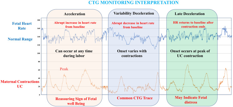
The computerization of both fetal heart rate (FHR) and intelligent classification modeling of the cardiotocograph (CTG) is one of the approaches that are utilized in assisting obstetricians in conducting initial interpretation based on (CTG) analysis. CTG tracing interpretation is crucial for the monitoring of the fetal status during weeks into the pregnancy and childbirth. Most contemporary studies rely on computer-assisted fetal heart rate (FHR) feature extraction and CTG categorization to determine the best precise diagnosis for tracking fetal health during pregnancy. Furthermore, through the utilization of a computer-assisted fetal monitoring system, the FHR patterns can be precisely detected and categorized.

OBJECTIVE

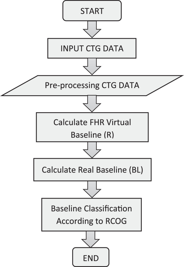
The goal of this project is to create a reliable feature extraction algorithm for the FHR as well as a systematic and viable classifier for the CTG through the utilization of the MATLAB platform, all the while adhering to the recognized Royal College of Obstetricians and Gynecologists (RCOG) recommendations.

METHOD

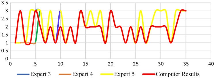
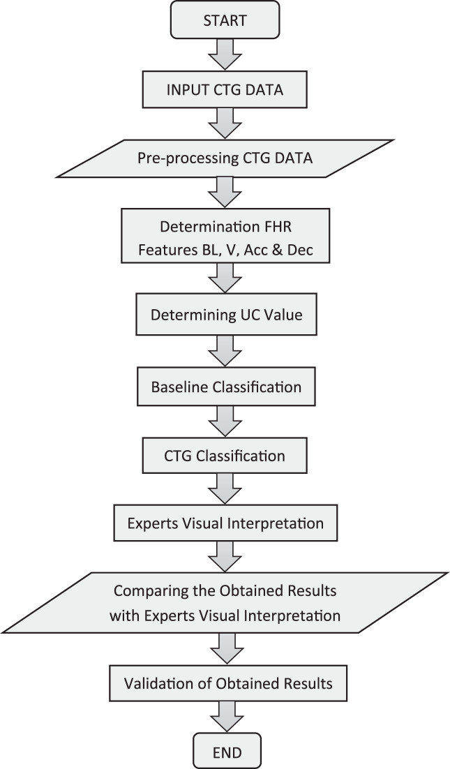
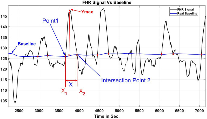
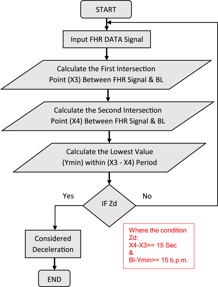
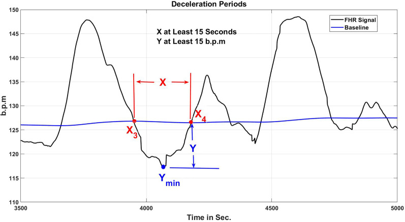
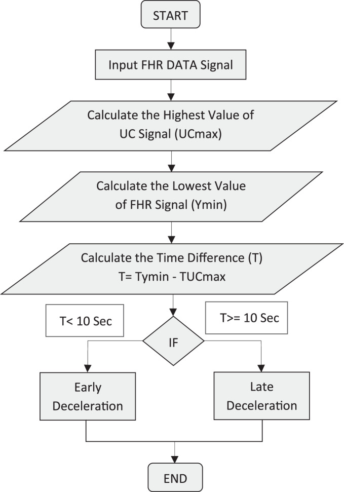
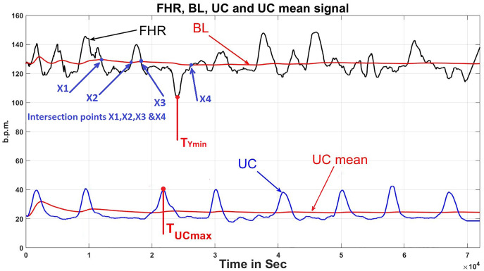
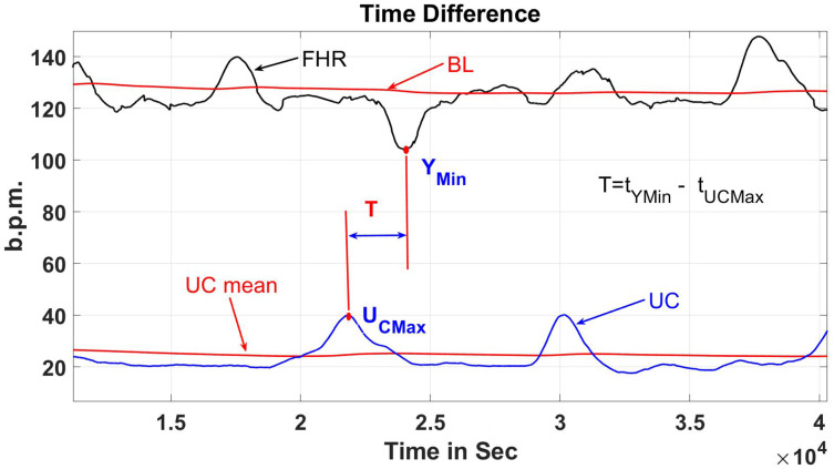
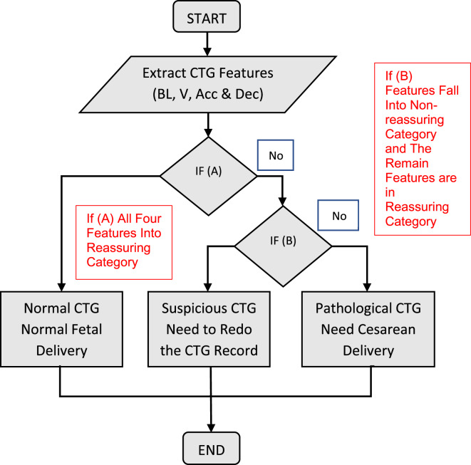
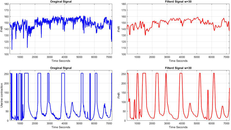
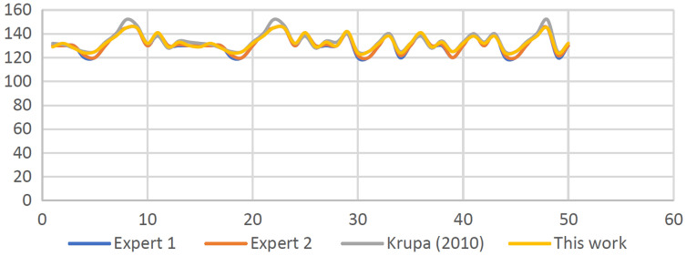
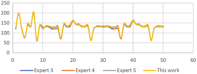
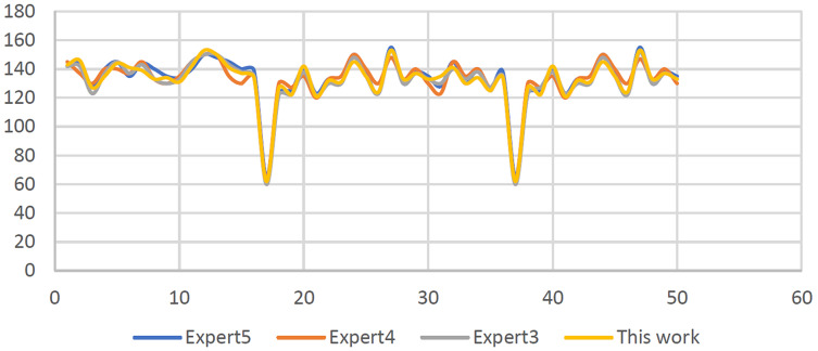
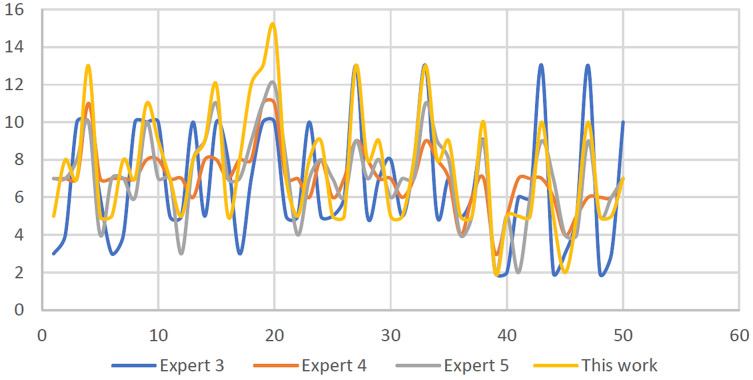
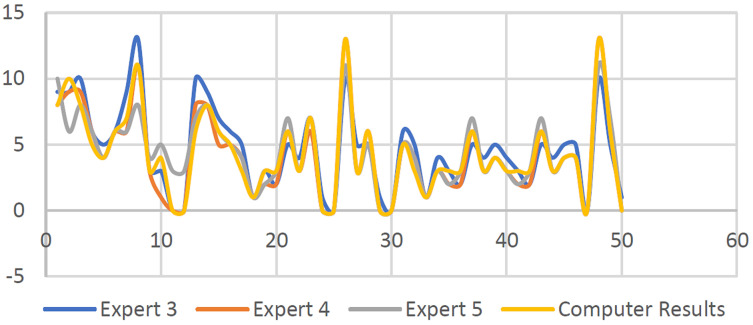
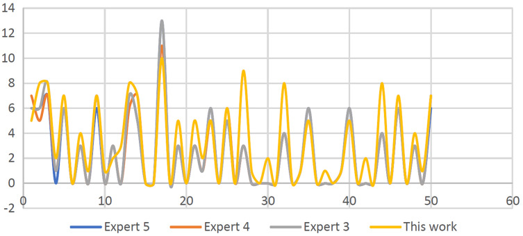
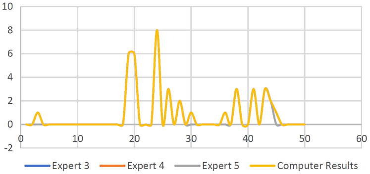
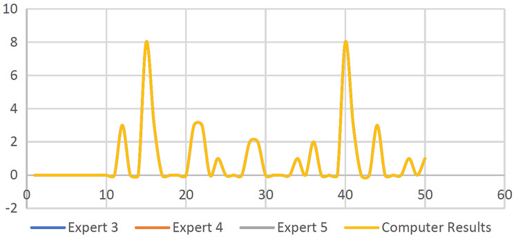
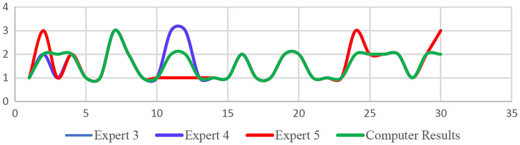
The compiled CTG data from spiky artifacts were cleaned by a specifically created application and compensated for missing data using the guidelines provided by RCOG and the MATLAB toolbox after the implemented data has been processed and the FHR fundamental features have been extracted, for example, the baseline, acceleration, deceleration, and baseline variability. This is followed by the classification phase based on the MATLAB environment. Next, using the guideline provided by the RCOG, the signals patterns of CTG were classified into three categories specifically as normal, abnormal (suspicious), or pathological. Furthermore, to ensure the effectiveness of the created computerized procedure and confirm the robustness of the method, the visual interpretation performed by five obstetricians is compared with the results utilizing the computerized version for the 150 CTG signals.

RESULTS

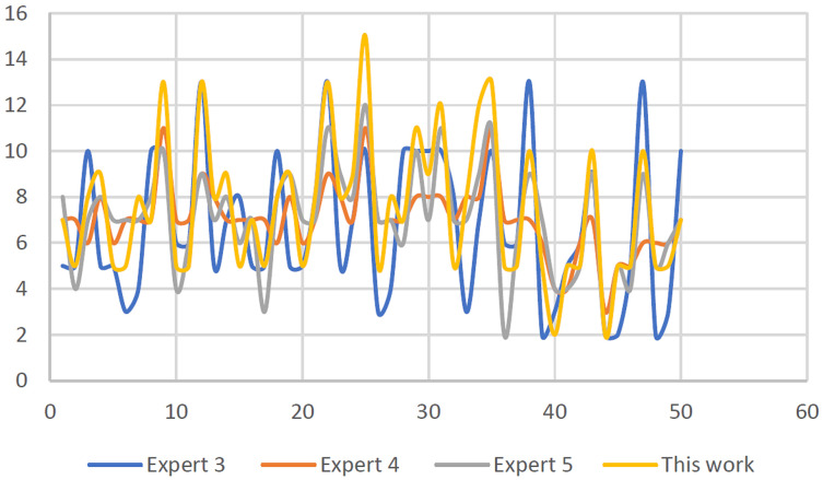
The attained CTG signal categorization results revealed that there is variability, particularly a trivial dissimilarity of approximately (+/-4 and 6) beats per minute (b.p.m.). It was demonstrated that obstetricians' observations coincide with algorithms based on deceleration type and number, except for acceleration values that differ by up to (+/-4).

DISCUSSION

The results obtained based on CTG interpretation showed that the utilization of the computerized approach employed in infirmaries and home care services for pregnant women is indeed suitable.

CONCLUSIONS

The classification based on CTG that was used for the interpretation of the FHR attribute as discussed in this study is based on the RCOG guidelines. The system is evaluated and validated by experts based on their expert opinions and was compared with the CTG feature extraction and classification algorithms developed using MATLAB.

摘要

背景

胎儿心率(FHR)的计算机化以及产程图(CTG)的智能分类建模是用于协助产科医生基于CTG分析进行初步解读的方法之一。CTG描记图解读对于孕期和分娩期间胎儿状况的监测至关重要。大多数当代研究依靠计算机辅助的胎儿心率(FHR)特征提取和CTG分类来确定孕期跟踪胎儿健康的最佳精确诊断。此外,通过使用计算机辅助胎儿监测系统,可以精确检测和分类FHR模式。

目的

本项目的目标是通过利用MATLAB平台创建一种可靠的FHR特征提取算法以及一种系统且可行的CTG分类器,同时遵循公认的皇家妇产科医师学院(RCOG)的建议。

方法

从尖峰伪影编译的CTG数据通过专门创建的应用程序进行清理,并在处理实施数据并提取FHR基本特征(例如基线、加速、减速和基线变异性)后,根据RCOG和MATLAB工具箱提供的指南对缺失数据进行补偿。接下来是基于MATLAB环境的分类阶段。然后,根据RCOG提供的指南,将CTG的信号模式具体分为三类,即正常、异常(可疑)或病理性。此外,为确保所创建的计算机化程序的有效性并确认该方法的稳健性,将五位产科医生进行的视觉解读与150个CTG信号的计算机化版本的结果进行比较。

结果

获得的CTG信号分类结果显示存在变异性,特别是每分钟约(±4和6)次心跳(b.p.m.)的微小差异。结果表明,除了加速值相差高达(±4)外,产科医生的观察结果与基于减速类型和数量的算法一致。

讨论

基于CTG解读获得的结果表明,在医院和孕妇家庭护理服务中采用的计算机化方法确实适用。

结论

本研究中用于解读FHR属性的基于CTG的分类是基于RCOG指南的。该系统由专家根据他们的专业意见进行评估和验证,并与使用MATLAB开发的CTG特征提取和分类算法进行比较。

https://cdn.ncbi.nlm.nih.gov/pmc/blobs/9989/9454876/ba75b1aeb1b1/peerj-cs-08-1050-g033.jpg
https://cdn.ncbi.nlm.nih.gov/pmc/blobs/9989/9454876/dfe46b25c44e/peerj-cs-08-1050-g001.jpg
https://cdn.ncbi.nlm.nih.gov/pmc/blobs/9989/9454876/1ad30a1e68cd/peerj-cs-08-1050-g002.jpg
https://cdn.ncbi.nlm.nih.gov/pmc/blobs/9989/9454876/f080bc5a2635/peerj-cs-08-1050-g003.jpg
https://cdn.ncbi.nlm.nih.gov/pmc/blobs/9989/9454876/fe8db08bc9da/peerj-cs-08-1050-g004.jpg
https://cdn.ncbi.nlm.nih.gov/pmc/blobs/9989/9454876/722e266d2675/peerj-cs-08-1050-g005.jpg
https://cdn.ncbi.nlm.nih.gov/pmc/blobs/9989/9454876/917f44cc1b8d/peerj-cs-08-1050-g006.jpg
https://cdn.ncbi.nlm.nih.gov/pmc/blobs/9989/9454876/81aa5722cfd2/peerj-cs-08-1050-g007.jpg
https://cdn.ncbi.nlm.nih.gov/pmc/blobs/9989/9454876/c406aed78fa5/peerj-cs-08-1050-g008.jpg
https://cdn.ncbi.nlm.nih.gov/pmc/blobs/9989/9454876/bc70d3dccb48/peerj-cs-08-1050-g009.jpg
https://cdn.ncbi.nlm.nih.gov/pmc/blobs/9989/9454876/4faf85641309/peerj-cs-08-1050-g010.jpg
https://cdn.ncbi.nlm.nih.gov/pmc/blobs/9989/9454876/c07d62030128/peerj-cs-08-1050-g011.jpg
https://cdn.ncbi.nlm.nih.gov/pmc/blobs/9989/9454876/1f04b4d1eeb3/peerj-cs-08-1050-g012.jpg
https://cdn.ncbi.nlm.nih.gov/pmc/blobs/9989/9454876/868b7a6020e3/peerj-cs-08-1050-g013.jpg
https://cdn.ncbi.nlm.nih.gov/pmc/blobs/9989/9454876/cdfcfe5959b1/peerj-cs-08-1050-g014.jpg
https://cdn.ncbi.nlm.nih.gov/pmc/blobs/9989/9454876/6c6686569c7c/peerj-cs-08-1050-g015.jpg
https://cdn.ncbi.nlm.nih.gov/pmc/blobs/9989/9454876/2e5e6c8aeae8/peerj-cs-08-1050-g016.jpg
https://cdn.ncbi.nlm.nih.gov/pmc/blobs/9989/9454876/380d8f870d26/peerj-cs-08-1050-g017.jpg
https://cdn.ncbi.nlm.nih.gov/pmc/blobs/9989/9454876/69c7592e2cbd/peerj-cs-08-1050-g018.jpg
https://cdn.ncbi.nlm.nih.gov/pmc/blobs/9989/9454876/719923493432/peerj-cs-08-1050-g019.jpg
https://cdn.ncbi.nlm.nih.gov/pmc/blobs/9989/9454876/3dbfdf723f45/peerj-cs-08-1050-g020.jpg
https://cdn.ncbi.nlm.nih.gov/pmc/blobs/9989/9454876/d9c8d7c14123/peerj-cs-08-1050-g021.jpg
https://cdn.ncbi.nlm.nih.gov/pmc/blobs/9989/9454876/865336eae895/peerj-cs-08-1050-g022.jpg
https://cdn.ncbi.nlm.nih.gov/pmc/blobs/9989/9454876/b995c2dfc2e2/peerj-cs-08-1050-g023.jpg
https://cdn.ncbi.nlm.nih.gov/pmc/blobs/9989/9454876/b6aa1511cbbc/peerj-cs-08-1050-g024.jpg
https://cdn.ncbi.nlm.nih.gov/pmc/blobs/9989/9454876/9cc868c97d0f/peerj-cs-08-1050-g025.jpg
https://cdn.ncbi.nlm.nih.gov/pmc/blobs/9989/9454876/b761a3e006ca/peerj-cs-08-1050-g026.jpg
https://cdn.ncbi.nlm.nih.gov/pmc/blobs/9989/9454876/9d6e1373baaf/peerj-cs-08-1050-g027.jpg
https://cdn.ncbi.nlm.nih.gov/pmc/blobs/9989/9454876/e59595bdecb2/peerj-cs-08-1050-g028.jpg
https://cdn.ncbi.nlm.nih.gov/pmc/blobs/9989/9454876/371ec0dde1b1/peerj-cs-08-1050-g029.jpg
https://cdn.ncbi.nlm.nih.gov/pmc/blobs/9989/9454876/1f4960967e3a/peerj-cs-08-1050-g030.jpg
https://cdn.ncbi.nlm.nih.gov/pmc/blobs/9989/9454876/4c910b90a1cd/peerj-cs-08-1050-g031.jpg
https://cdn.ncbi.nlm.nih.gov/pmc/blobs/9989/9454876/de773468323d/peerj-cs-08-1050-g032.jpg
https://cdn.ncbi.nlm.nih.gov/pmc/blobs/9989/9454876/ba75b1aeb1b1/peerj-cs-08-1050-g033.jpg

相似文献

1
Intrapartum cardiotocography trace pattern pre-processing, features extraction and fetal health condition diagnoses based on RCOG guideline.基于英国皇家妇产科医师学院(RCOG)指南的产时胎心监护图模式预处理、特征提取及胎儿健康状况诊断
PeerJ Comput Sci. 2022 Aug 18;8:e1050. doi: 10.7717/peerj-cs.1050. eCollection 2022.
2
eCTG: an automatic procedure to extract digital cardiotocographic signals from digital images.eCTG:一种从数字图像中提取数字化胎心监护信号的自动程序。
Comput Methods Programs Biomed. 2018 Mar;156:133-139. doi: 10.1016/j.cmpb.2017.12.030. Epub 2018 Jan 2.
3
A systematic review of automated pre-processing, feature extraction and classification of cardiotocography.对胎心监护自动预处理、特征提取与分类的系统评价。
PeerJ Comput Sci. 2021 Apr 27;7:e452. doi: 10.7717/peerj-cs.452. eCollection 2021.
4
Developments in CTG analysis.产程图分析的进展
Baillieres Clin Obstet Gynaecol. 1996 Jun;10(2):185-209. doi: 10.1016/s0950-3552(96)80033-2.
5
Intelligent antepartum fetal monitoring via deep learning and fusion of cardiotocographic signals and clinical data.通过深度学习以及胎心监护信号与临床数据融合实现智能产前胎儿监测。
Health Inf Sci Syst. 2023 Mar 19;11(1):16. doi: 10.1007/s13755-023-00219-w. eCollection 2023 Dec.
6
Comparison of a novel computerized analysis program and visual interpretation of cardiotocography.一种新型计算机分析程序与胎心监护图视觉解读的比较。
PLoS One. 2014 Dec 1;9(12):e112296. doi: 10.1371/journal.pone.0112296. eCollection 2014.
7
[Relationship between computerized cardiotocography and perinatal outcomes].[计算机化胎心监护与围产期结局的关系]
Zhonghua Fu Chan Ke Za Zhi. 2001 Oct;36(10):581-4.
8
Critical Imperative for the Reform of British Interpretation of Fetal Heart Rate Decelerations: Analysis of FIGO and NICE Guidelines, Post-Truth Foundations, Cognitive Fallacies, Myths and Occam's Razor.英国胎儿心率减速解读改革的关键必要性:对国际妇产科联盟(FIGO)和英国国家卫生与临床优化研究所(NICE)指南、后真相基础、认知谬误、神话及奥卡姆剃刀原理的分析
J Clin Med Res. 2017 Apr;9(4):253-265. doi: 10.14740/jocmr2877e. Epub 2017 Feb 21.
9
The application of empirical mode decomposition for the enhancement of cardiotocograph signals.经验模态分解在增强胎心监护信号中的应用。
Physiol Meas. 2009 Aug;30(8):729-43. doi: 10.1088/0967-3334/30/8/001. Epub 2009 Jun 24.
10
Cardiotocography analysis by empirical dynamic modeling and Gaussian processes.基于经验动态建模和高斯过程的胎心监护分析
Front Bioeng Biotechnol. 2023 Jan 12;10:1057807. doi: 10.3389/fbioe.2022.1057807. eCollection 2022.

本文引用的文献

1
Systematic Review of Intrapartum Fetal Heart Rate Spectral Analysis and an Application in the Detection of Fetal Acidemia.产时胎儿心率频谱分析的系统评价及其在胎儿酸血症检测中的应用
Front Pediatr. 2021 Aug 2;9:661400. doi: 10.3389/fped.2021.661400. eCollection 2021.
2
A systematic review of automated pre-processing, feature extraction and classification of cardiotocography.对胎心监护自动预处理、特征提取与分类的系统评价。
PeerJ Comput Sci. 2021 Apr 27;7:e452. doi: 10.7717/peerj-cs.452. eCollection 2021.
3
Fetal Heart Rate Variability Is Affected by Fetal Movements: A Systematic Review.
胎儿心率变异性受胎动影响:一项系统评价。
Front Physiol. 2020 Sep 30;11:578898. doi: 10.3389/fphys.2020.578898. eCollection 2020.
4
Heart Rate Variability in the Perinatal Period: A Critical and Conceptual Review.围产期心率变异性:一项批判性和概念性综述。
Front Neurosci. 2020 Sep 25;14:561186. doi: 10.3389/fnins.2020.561186. eCollection 2020.
5
Relative accuracy of computerized intrapartum fetal heart rate pattern recognition by ultrasound and abdominal electrocardiogram detection.超声和腹部心电图检测计算机识别产时胎儿心率图形的相对准确性。
Acta Obstet Gynecol Scand. 2020 Mar;99(3):413-422. doi: 10.1111/aogs.13760. Epub 2019 Dec 9.
6
Integrating machine learning techniques and physiology based heart rate features for antepartum fetal monitoring.整合机器学习技术和基于生理学的心率特征进行产前胎儿监测。
Comput Methods Programs Biomed. 2020 Mar;185:105015. doi: 10.1016/j.cmpb.2019.105015. Epub 2019 Oct 17.
7
Prediction of intrapartum fetal hypoxia considering feature selection algorithms and machine learning models.考虑特征选择算法和机器学习模型的产时胎儿缺氧预测
Health Inf Sci Syst. 2019 Aug 20;7(1):17. doi: 10.1007/s13755-019-0079-z. eCollection 2019 Dec.
8
Fetal cardiotocography monitoring using Legendre neural networks.使用勒让德神经网络进行胎儿心率宫缩图监测。
Biomed Tech (Berl). 2019 Dec 18;64(6):669-675. doi: 10.1515/bmt-2018-0074.
9
Computer-Aided Diagnosis System of Fetal Hypoxia Incorporating Recurrence Plot With Convolutional Neural Network.结合递归图与卷积神经网络的胎儿缺氧计算机辅助诊断系统
Front Physiol. 2019 Mar 12;10:255. doi: 10.3389/fphys.2019.00255. eCollection 2019.
10
Prognostic model based on image-based time-frequency features and genetic algorithm for fetal hypoxia assessment.基于图像时频特征和遗传算法的胎儿缺氧评估预后模型。
Comput Biol Med. 2018 Aug 1;99:85-97. doi: 10.1016/j.compbiomed.2018.06.003. Epub 2018 Jun 6.