Department of Pathology and Microbiology, University of Nebraska Medical Center, Nebraska Medical Center, Omaha, Nebraska.
Department of Microbiology, Immunology, and Biochemistry, University of Tennessee Health Science Center, Memphis, Tennessee.
PLoS Pathog. 2022 Sep 12;18(9):e1010836. doi: 10.1371/journal.ppat.1010836. eCollection 2022 Sep.
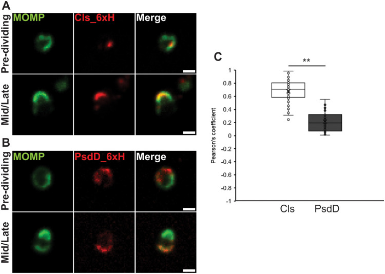
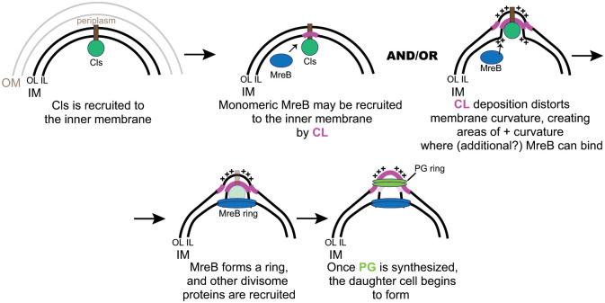
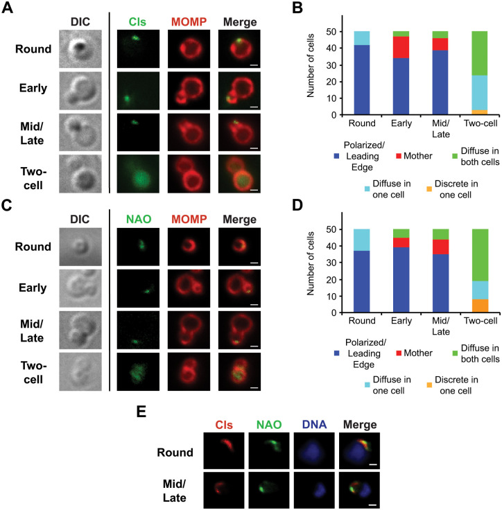
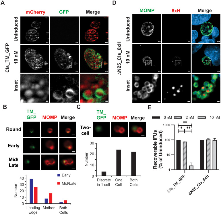
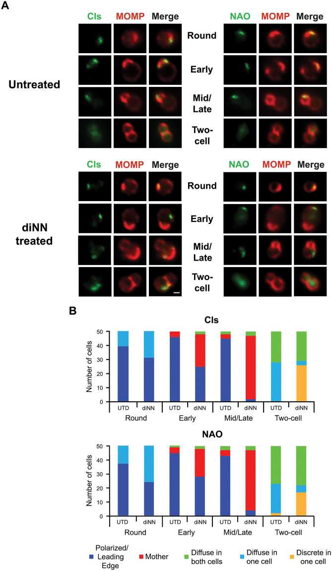
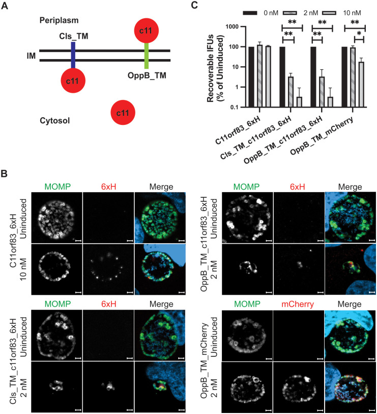
Pathogenic Chlamydia species are coccoid bacteria that use the rod-shape determining protein MreB to direct septal peptidoglycan synthesis during their polarized cell division process. How the site of polarized budding is determined in this bacterium, where contextual features like membrane curvature are seemingly identical, is unclear. We hypothesized that the accumulation of the phospholipid, cardiolipin (CL), in specific regions of the cell membrane induces localized membrane changes that trigger the recruitment of MreB to the site where the bud will arise. To test this, we ectopically expressed cardiolipin synthase (Cls) and observed a polar distribution for this enzyme in Chlamydia trachomatis. In early division intermediates, Cls was restricted to the bud site where MreB is localized and peptidoglycan synthesis is initiated. The localization profile of 6xHis tagged Cls (Cls_6xH) throughout division mimicked the distribution of lipids that stain with NAO, a dye that labels CL. Treatment of Chlamydia with 3',6-dinonylneamine (diNN), an antibiotic targeting CL-containing membrane domains, resulted in redistribution of Cls_6xH and NAO-staining phospholipids. In addition, 6xHis tagged MreB localization was altered by diNN treatment, suggesting an upstream regulatory role for CL-containing membranes in directing the assembly of MreB. This hypothesis is consistent with the observation that the clustered localization of Cls_6xH is not dependent upon MreB function or peptidoglycan synthesis. Furthermore, expression of a CL-binding protein at the inner membrane of C. trachomatis dramatically inhibited bacterial growth supporting the importance of CL in the division process. Our findings implicate a critical role for localized CL synthesis in driving MreB assembly at the bud site during the polarized cell division of Chlamydia.
致病衣原体是球形细菌,它们在极性细胞分裂过程中使用棒状决定蛋白 MreB 来指导隔膜肽聚糖的合成。在这种细菌中,如何确定极性出芽的部位,在那里膜曲率等上下文特征似乎是相同的,目前还不清楚。我们假设,磷脂心磷脂(CL)在细胞膜的特定区域积累会引起局部膜变化,从而触发 MreB 募集到将要出芽的部位。为了验证这一点,我们异位表达了心磷脂合酶(Cls),并观察到 Chlamydia trachomatis 中这种酶的极性分布。在早期分裂中间体中,Cls 局限于芽的部位,MreB 定位于该处,肽聚糖合成开始。6xHis 标记的 Cls(Cls_6xH)在整个分裂过程中的定位模式类似于用 NAO 染色的脂质的分布,NAO 是一种标记 CL 的染料。用 3',6-二壬基胺(diNN)处理衣原体,一种针对含有 CL 的膜结构域的抗生素,导致 Cls_6xH 和 NAO 染色的磷脂重新分布。此外,diNN 处理改变了 6xHis 标记的 MreB 的定位,表明含有 CL 的膜在指导 MreB 组装中起上游调节作用。这一假设与以下观察结果一致,即 Cls_6xH 的聚类定位不依赖于 MreB 功能或肽聚糖合成。此外,在沙眼衣原体的内膜上表达 CL 结合蛋白,极大地抑制了细菌的生长,这支持了 CL 在分裂过程中的重要性。我们的发现表明,在衣原体的极性细胞分裂过程中,局部 CL 合成在驱动 MreB 组装到芽部位方面起着关键作用。