Kyi Priscilla, Hendee Kathryn, Hunyenyiwa Tendai, Matus Kienna, Mammoto Tadanori, Mammoto Akiko
Department of Pediatrics, Medical College of Wisconsin, Milwaukee, WI, United States.
Department of Cell Biology, Neurobiology and Anatomy, Medical College of Wisconsin, Milwaukee, WI, United States.
Front Med (Lausanne). 2022 Sep 20;9:908639. doi: 10.3389/fmed.2022.908639. eCollection 2022.
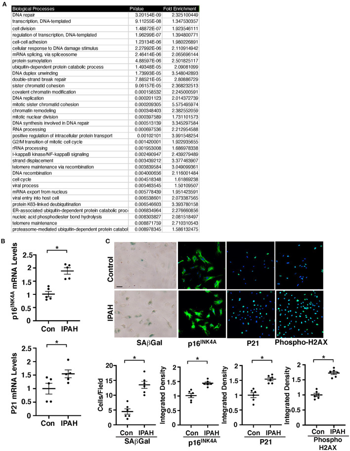
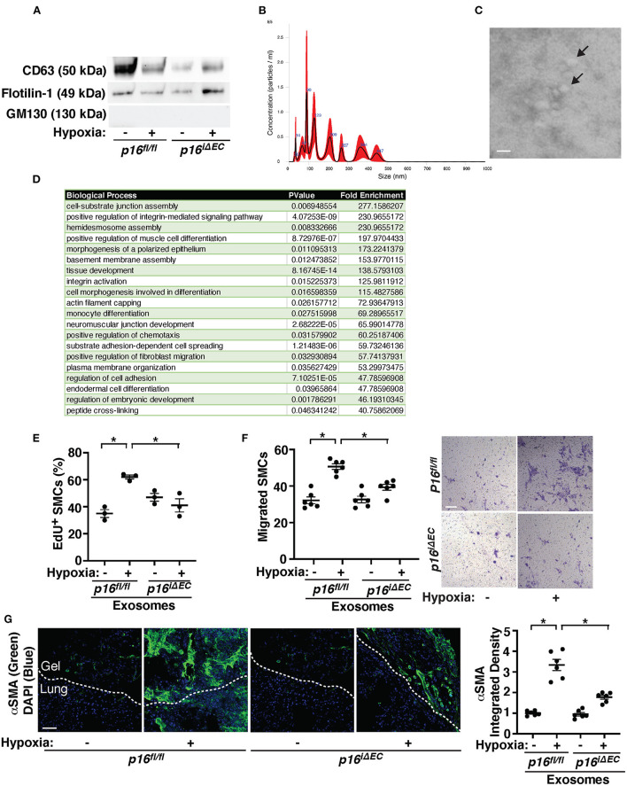
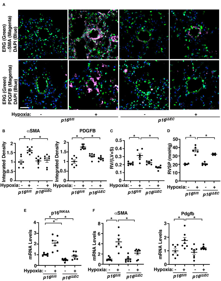
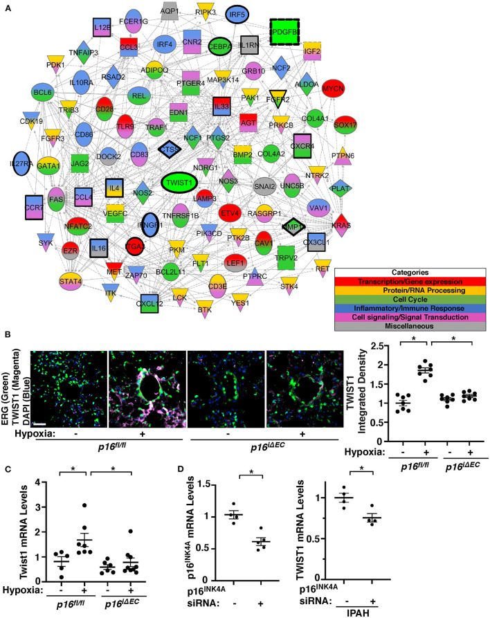
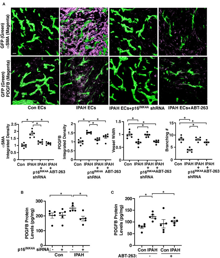
Uncontrolled accumulation of pulmonary artery smooth muscle cells (PASMCs) to the distal pulmonary arterioles (PAs) is one of the major characteristics of pulmonary hypertension (PH). Cellular senescence contributes to aging and lung diseases associated with PH and links to PH progression. However, the mechanism by which cellular senescence controls vascular remodeling in PH is not fully understood. The levels of senescence marker, p16 and senescence-associated β-galactosidase (SA-β-gal) activity are higher in PA endothelial cells (ECs) isolated from idiopathic pulmonary arterial hypertension (IPAH) patients compared to those from healthy individuals. Hypoxia-induced accumulation of α-smooth muscle actin (αSMA)-positive cells to the PAs is attenuated in ( ) mice after tamoxifen induction. We have reported that endothelial TWIST1 mediates hypoxia-induced vascular remodeling by increasing platelet-derived growth factor (PDGFB) expression. Transcriptomic analyses of IPAH patient lungs or hypoxia-induced mouse lung ECs reveal the alteration of senescence-related gene expression and their interaction with TWIST1. Knockdown of p16 attenuates the expression of PDGFB and TWIST1 in IPAH patient PAECs or hypoxia-treated mouse lungs and suppresses accumulation of αSMA-positive cells to the supplemented ECs in the gel implanted on the mouse lungs. Hypoxia-treated mouse lung EC-derived exosomes stimulate DNA synthesis and migration of PASMCs and in the gel implanted on the mouse lungs, while mouse lung EC-derived exosomes inhibit the effects. These results suggest that endothelial senescence modulates TWIST1-PDGFB signaling and controls vascular remodeling in PH.
肺动脉平滑肌细胞(PASMCs)在远端肺小动脉(PAs)中不受控制的积累是肺动脉高压(PH)的主要特征之一。细胞衰老导致与PH相关的衰老和肺部疾病,并与PH的进展有关。然而,细胞衰老控制PH中血管重塑的机制尚未完全了解。与健康个体相比,从特发性肺动脉高压(IPAH)患者分离的肺动脉内皮细胞(ECs)中衰老标志物p16的水平和衰老相关β-半乳糖苷酶(SA-β-gal)活性更高。他莫昔芬诱导后,( )小鼠中缺氧诱导的α-平滑肌肌动蛋白(αSMA)阳性细胞在PAs中的积累减弱。我们报道过内皮细胞TWIST1通过增加血小板衍生生长因子(PDGFB)的表达介导缺氧诱导的血管重塑。对IPAH患者肺组织或缺氧诱导的小鼠肺ECs进行转录组分析,揭示了衰老相关基因表达的改变及其与TWIST1的相互作用。敲低p16可减弱IPAH患者肺动脉内皮细胞(PAECs)或缺氧处理的小鼠肺组织中PDGFB和TWIST1的表达,并抑制αSMA阳性细胞在植入小鼠肺组织的凝胶中对补充的ECs的积累。缺氧处理的小鼠肺ECs衍生的外泌体刺激PASMCs的DNA合成和迁移,而在植入小鼠肺组织的凝胶中,( )小鼠肺ECs衍生的外泌体则抑制这种作用。这些结果表明,内皮细胞衰老调节TWIST1-PDGFB信号通路并控制PH中的血管重塑。