Department of Bioscience Technology, Chung Yuan Christian University, Chung Li District, Taoyuan City 32023, Taiwan.
Department of Radiation Oncology, Mackay Memorial Hospital, Taipei 104, Taiwan.
Int J Mol Sci. 2022 Nov 2;23(21):13389. doi: 10.3390/ijms232113389.
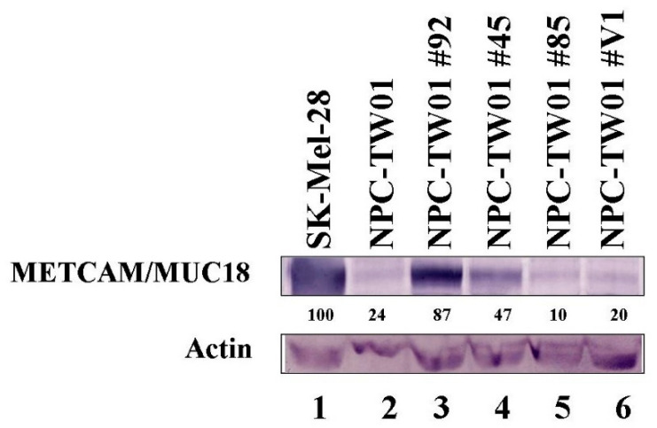
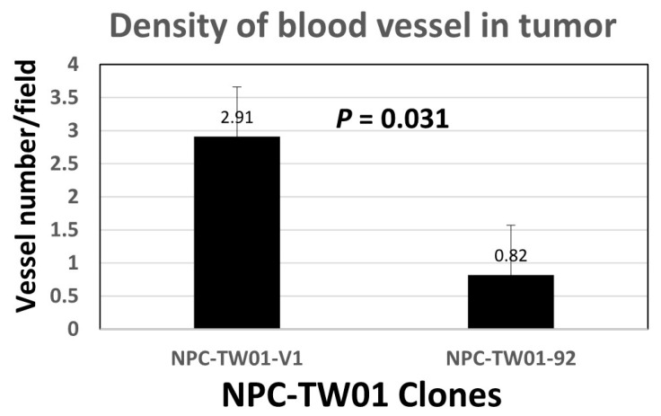
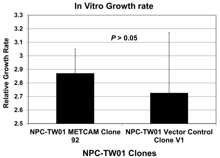
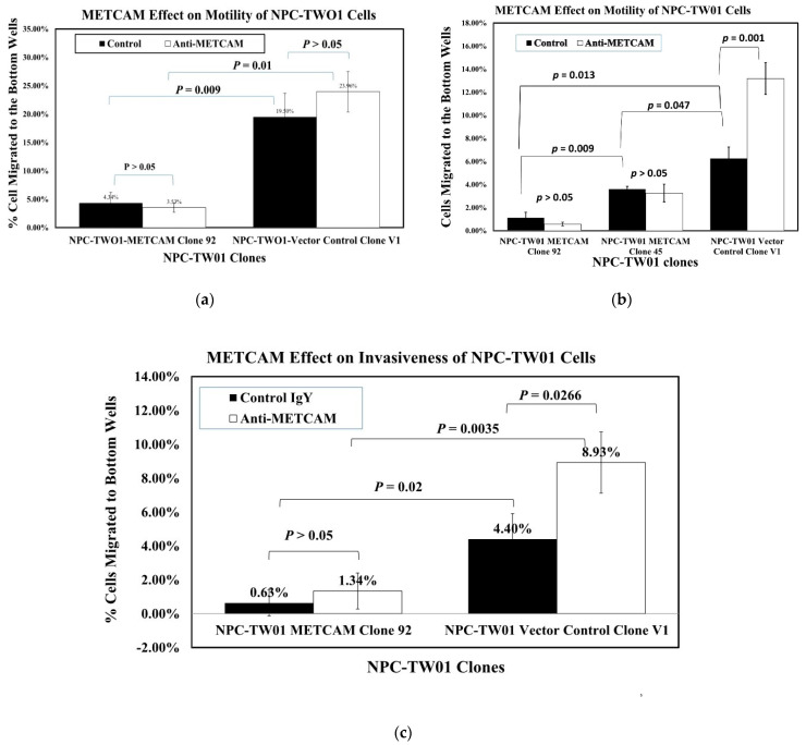
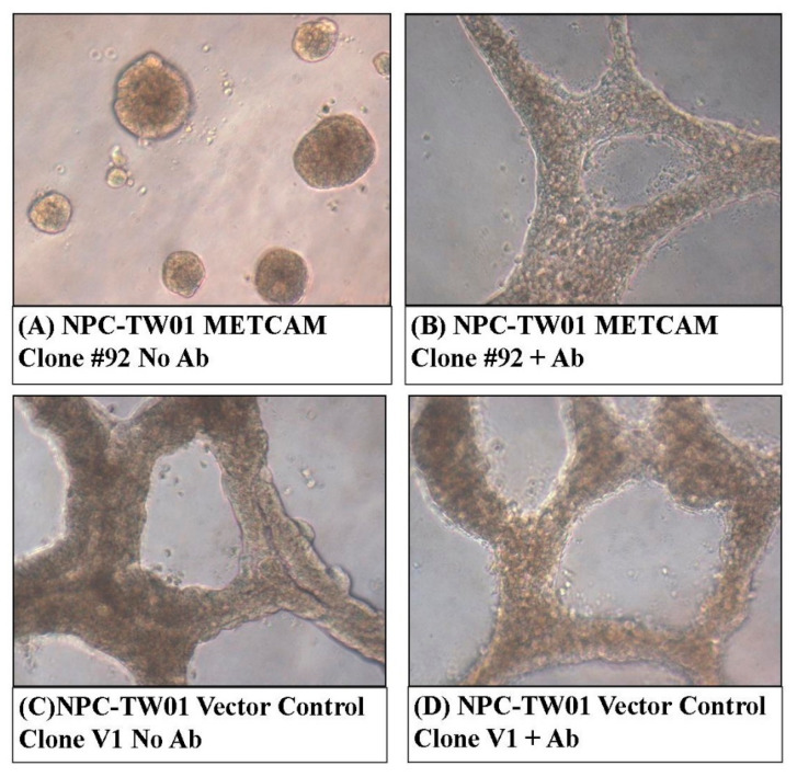
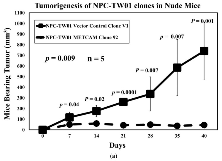
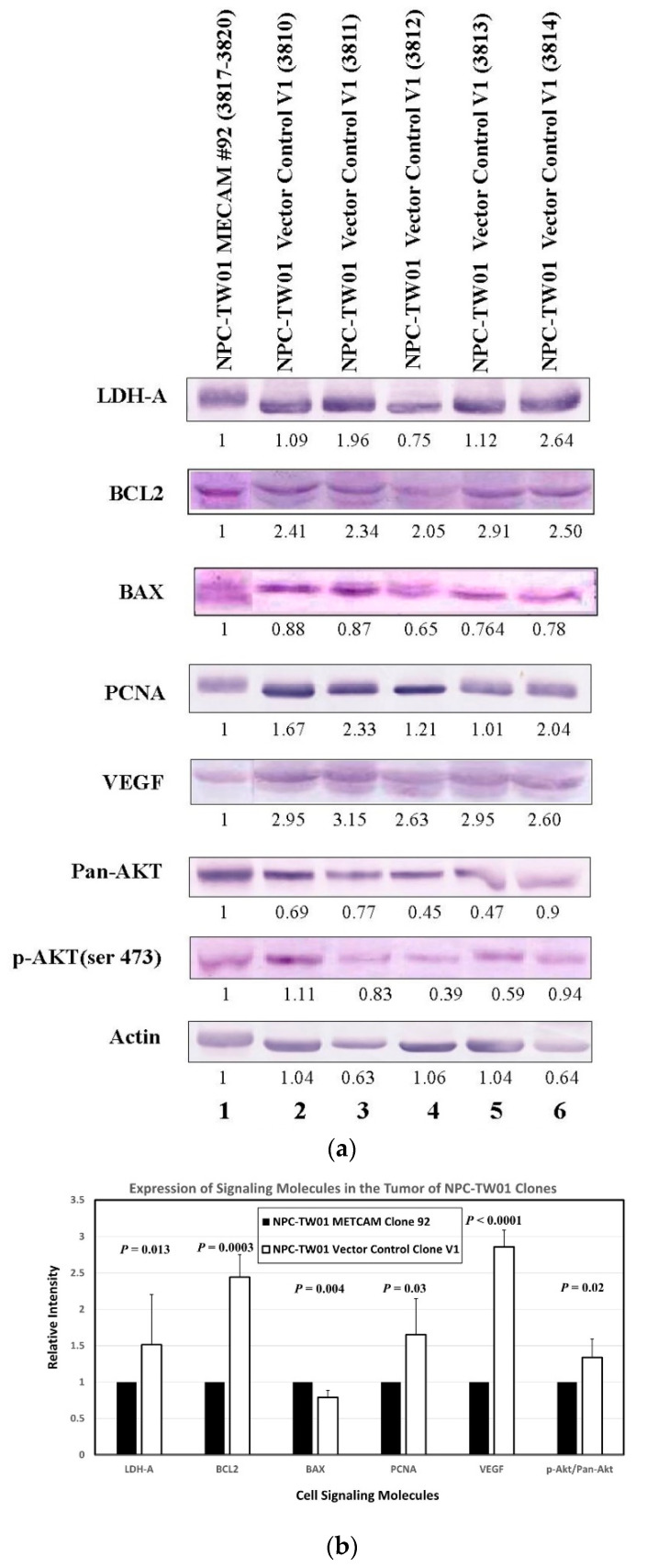
From previous studies of negatively correlating the expression of human METCAM/MUC18 with the pathology of nasopharyngeal carcinoma (NPC), we have suggested that human METCAM/MUC18 (huMETCAM/MUC18) might play a tumor suppressor role in the development of nasopharyngeal carcinoma. To scrutinize this hypothesis, we investigated the effects of huMETCAM/MUC18's over-expression on in vitro cellular behavior and on the in vivo tumorigenesis of one NPC cell line (NPC-TW01). HuMETCAM/MUC18 cDNA was first transfected into the NPC-TW01 cell line, which was established from NPC type I, and many G418-resistant clones were obtained. Then, two NPC-TW01 clones, which expressed high and medium levels of huMETCAM/MUC18, respectively, and one empty vector (control) clone were used to test the effects of huMETCAM/MUC18's over-expression on in vitro behaviors and on in vivo tumorigenesis (via subcutaneous injection) in athymic nude mice (Balb/cAnN.Cg-/Cr1Nar1). The time course of tumor proliferation and the final tumor weights were determined. Tumor sections were used for the histology and immunohistochemistry (IHC) studies. Tumor lysates were used for determining the expression levels of huMETCAM/MUC18 and various downstream key effectors. HuMETCAM/MUC18's over-expression reduced in vitro motility and invasiveness and altered growth behaviors in 3D basement membrane culture assays, and it decreased the in vivo tumorigenicity of the NPC-TW01 cells. The tumor cells from a high-expressing clone were clustered and confined in small areas, whereas those from a vector control clone were more spread out, suggesting that the tumor cells from the high-expressing clone appeared to stay dormant in micro-clusters. Expression levels of the proliferation index, an index of the metabolic switch to aerobic glycolysis, angiogenesis indexes, and survival pathway indexes were reduced, whereas the pro-apoptosis index increased in the corresponding tumors. The over-expression of huMETCAM/MUC18 in the NPC-TW01 cells decreased the epithelial-to-mesenchymal transition and the in vitro and in vitro tumorigenesis, suggesting that it plays a tumor suppressor role in the development of type I NPC, perhaps by increasing apoptosis and decreasing angiogenesis, proliferation, and the metabolic switch to aerobic glycolysis.
从之前研究表明人类 METCAM/MUC18 的表达与鼻咽癌(NPC)的病理呈负相关,我们推测人类 METCAM/MUC18(huMETCAM/MUC18)可能在鼻咽癌的发展中发挥肿瘤抑制作用。为了验证这一假设,我们研究了 huMETCAM/MUC18 的过表达对一种 NPC 细胞系(NPC-TW01)的体外细胞行为和体内致瘤性的影响。首先将 huMETCAM/MUC18 cDNA 转染到 NPC-TW01 细胞系中,该细胞系源自 I 型 NPC,获得了许多 G418 抗性克隆。然后,使用两种分别表达高和中水平 huMETCAM/MUC18 的 NPC-TW01 克隆和一个空载体(对照)克隆,通过皮下注射在裸鼠(Balb/cAnN.Cg-/Cr1Nar1)中测试 huMETCAM/MUC18 过表达对体外行为和体内致瘤性(通过皮下注射)的影响。确定肿瘤增殖的时间过程和最终肿瘤重量。使用肿瘤切片进行组织学和免疫组织化学(IHC)研究。使用肿瘤裂解物测定 huMETCAM/MUC18 和各种下游关键效应物的表达水平。huMETCAM/MUC18 的过表达降低了体外运动性和侵袭性,并改变了 3D 基底膜培养测定中的生长行为,降低了 NPC-TW01 细胞的体内致瘤性。高表达克隆的肿瘤细胞聚集并局限在小区域,而载体对照克隆的肿瘤细胞则更加扩散,这表明高表达克隆的肿瘤细胞似乎在微簇中处于休眠状态。增殖指数(有氧糖酵解代谢转换的指标)、血管生成指数和生存途径指数的表达水平降低,而促凋亡指数增加。在 NPC-TW01 细胞中过表达 huMETCAM/MUC18 降低了上皮-间充质转化以及体外和体内致瘤性,这表明它在 I 型 NPC 的发展中发挥肿瘤抑制作用,可能通过增加凋亡、减少血管生成、增殖和有氧糖酵解的代谢转换来实现。