Urology Department, Hospital Senhora da Oliveira, Creixomil, Portugal.
Tumour and Microenvironment Interactions Group, i3S, Instituto de Investigação e Inovação Em Saúde da Universidade Do Porto, Rua Alfredo Allen, 208, 4200-135, Porto, Portugal.
Sci Rep. 2022 Nov 19;12(1):19956. doi: 10.1038/s41598-022-24418-9.
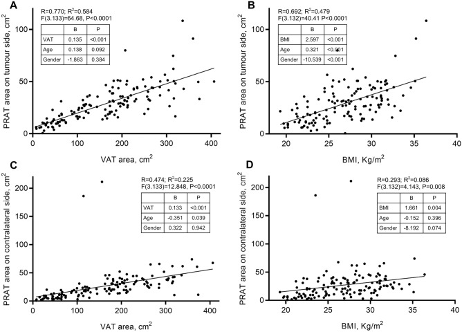
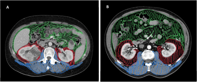
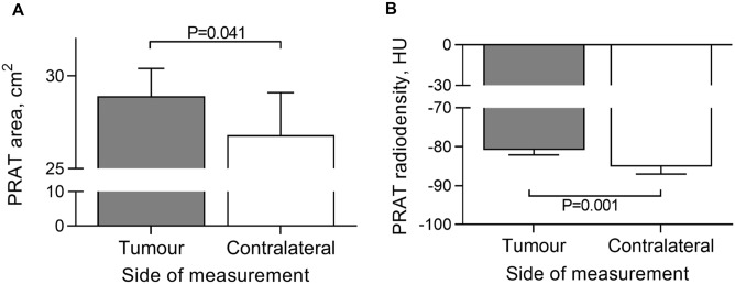
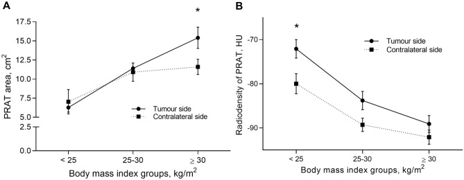
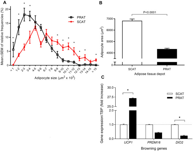
Increasing evidence supports a role for local fat depots in cancer outcomes. Despite the robust positive association of obesity with renal cell carcinoma (RCCa) diagnosis, increased adiposity is inversely related to RCCa oncological outcomes. Here, we sought to ascertain whether imagiologically assessed local fat depots associate with RCCa progression and survival and account for this apparent paradox. A retrospective cohort of renal carcinoma patients elective for nephrectomy (n = 137) were included. Beyond baseline clinicopathological characteristics, computed tomography (CT)-scans at the level of renal hilum evaluated areas and densities of different adipose tissue depots (perirenal, subcutaneous, visceral) and skeletal muscle (erector spinae, psoas and quadratus lumborum muscles) were analyzed. Univariate and multivariable Cox proportional hazards models were estimated following empirical analysis using stepwise Cox regression. Age, visceral adipose tissue (VAT) area and body mass index (BMI) predicted tumour-sided perirenal fat area (R = 0.584), which presented upregulated UCP1 expression by 27-fold (P = 0.026) and smaller adipocyte areas, compared with subcutaneous depot. Multivariate analyses revealed that increased area of perirenal adipose tissue (PRAT) on the contralateral and tumour side associate with improved progression-free survival (HR = 0.3, 95CI = 0.1-0.8, P = 0.019) and overall survival (HR = 0.3, 95CI = 0.1-0.7, P = 0.009). PRAT measurements using CT, might become a possible tool, well correlated with other measures of obesity such as VAT and BMI, that will improve determination of obesity and contribute to assess the risk for disease progression and mortality in renal cancer patients. Present data supports the obesity paradox in RCCa, assumed that larger PRAT areas seem to protect from disease progression and death.
越来越多的证据表明局部脂肪沉积在癌症结局中起作用。尽管肥胖与肾细胞癌(RCCa)诊断之间存在显著的正相关关系,但肥胖程度的增加与 RCCa 的肿瘤学结果呈负相关。在这里,我们试图确定影像学评估的局部脂肪沉积是否与 RCCa 的进展和生存相关,并解释这种明显的悖论。我们纳入了一组接受肾切除术的肾癌患者的回顾性队列(n=137)。除了基线临床病理特征外,还对肾门水平的计算机断层扫描(CT)评估了不同脂肪组织沉积(肾周、皮下、内脏)和骨骼肌(竖脊肌、腰大肌和腰方肌)的面积和密度。使用逐步 Cox 回归的经验分析估计了单变量和多变量 Cox 比例风险模型。年龄、内脏脂肪组织(VAT)面积和体重指数(BMI)预测肿瘤侧肾周脂肪面积(R=0.584),与皮下沉积相比,肿瘤侧肾周脂肪的 UCP1 表达上调了 27 倍(P=0.026),脂肪细胞面积更小。多变量分析显示,对侧和肿瘤侧肾周脂肪组织(PRAT)面积的增加与无进展生存期(HR=0.3,95%CI=0.1-0.8,P=0.019)和总生存期(HR=0.3,95%CI=0.1-0.7,P=0.009)的改善相关。使用 CT 测量 PRAT 可能成为一种可能的工具,与 VAT 和 BMI 等其他肥胖测量方法密切相关,这将有助于确定肥胖,并有助于评估肾癌患者疾病进展和死亡的风险。目前的数据支持 RCCa 中的肥胖悖论,即较大的 PRAT 面积似乎可以防止疾病进展和死亡。