de la Pinta Carolina, LaTorre Raquel García, Martínez-Lorca Alberto, Fernández Eva, Hernanz Raul, Martín Mercedes, Domínguez Jose A, Muñóz Teresa, Canales Elena, Vallejo Carmen, Alarza Marina, Hervás Asunción, Garví Manuel, Pino Vanesa, Sancho Sonsoles
Department of Radiation Oncology, Ramón y Cajal University Hospital, IRYCIS, Madrid, Spain.
Department of Radiology, Ramón y Cajal University Hospital, IRYCIS, Madrid, Spain.
J Clin Transl Res. 2022 Oct 22;8(6):465-469. eCollection 2022 Dec 29.
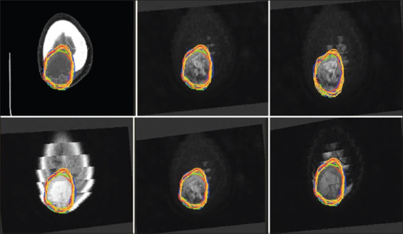
The optimal imaging test for gross tumor volume (GTV) delineation in non-spine bone metastases has not been defined. The use of stereotactic body radiotherapy (SBRT) requires accurate target delineation. Magnetic resonance imaging (MRI) and/or fludesoxyglucose positron emission tomography (18FDG-PET) allow for better visualization of the extent of bone metastases and optimizes the accuracy of tumor delineation for stereotactic radiotherapy compared to computed tomography (CT) alone. We evaluated the interobserver agreement in GTV of non-spine bone metastases in a single center and compared MRI and/or 18FDG-PET and CT in GTV delineation.
Anonymous CT and MRI and/or 18FDG-PET obtained from 10 non-spine bone metastases were analyzed by six radiation oncologists at our center. Images acquired by CT and MRI and/or 18FDG-PET were used to delineate 10 GTVs of non-spine bone metastases in the pelvis, extremities, and skull. The cases showed different characteristics: blastic and lytic metastases, and different primary cancers (lung, breast, prostate, rectum, urothelial, and biliary). In both CT and MRI and/or 18FDG-PET, the GTV volumes were compared. The index of agreement was evaluated according to Landis and Koch protocol.
The GTV volume as defined on MRI was in all cases larger or at least as large as the GTV volume on CT (=0.25). The median GTV volume on MRI was 3.15 cc (0.027-70.64 cc) compared to 2.8 cc on CT (0.075-77.95 cc). Interobserver variance and standard deviation were lower in CT than MRI (576.3 vs. 722.2 and 24.0 vs. 26.9, respectively). The level of agreement was fair (kappa=0.36) between CT and MRI. The median GTV volume on 18FDG-PET in five patients was 5.8 cc (0.46-64.17 cc), compared to 4.1 cc on CT (0.99-54.2 cc) (=0.236). Interobserver variance and standard deviation in CT, MRI, and 18FDG-PET were 576.3 versus 722.2 versus 730.5 and 24 versus 26.9 versus 27.0, respectively. The level of agreement was slight (kappa=0.08) between CT and 18FDG-PET.
Interobserver variance in non-spine bone metastases was equal when MRI and PET were compared to CT. CT was associated with the lowest variance and standard deviation. Compared to CT GTVs, the GTVs rendered from MRI images had fair agreement, while the GTVs rendered from 18FDG-PET had only slight agreement.
The delimitation of the treatment volume in non-spine bone metastases with SBRT is important for the results determining its efficacy. It is therefore essential to know the variability and to manage it to achieve the highest quality of treatment.
尚未确定用于非脊柱骨转移瘤大体肿瘤体积(GTV)勾画的最佳影像学检查方法。立体定向体部放疗(SBRT)的应用需要精确的靶区勾画。与单纯计算机断层扫描(CT)相比,磁共振成像(MRI)和/或氟脱氧葡萄糖正电子发射断层扫描(18FDG-PET)能更好地显示骨转移瘤的范围,并优化立体定向放疗中肿瘤勾画的准确性。我们评估了单中心非脊柱骨转移瘤GTV的观察者间一致性,并比较了MRI和/或18FDG-PET与CT在GTV勾画中的差异。
我们中心的6名放射肿瘤学家分析了从10例非脊柱骨转移瘤患者获取的匿名CT、MRI和/或18FDG-PET图像。利用CT、MRI和/或18FDG-PET获取的图像勾画10例骨盆、四肢和颅骨非脊柱骨转移瘤的GTV。这些病例具有不同特征:成骨和溶骨性转移,以及不同的原发癌(肺癌、乳腺癌、前列腺癌、直肠癌、尿路上皮癌和胆管癌)。比较CT、MRI和/或18FDG-PET中GTV的体积。根据Landis和Koch方案评估一致性指数。
MRI定义的GTV体积在所有病例中均大于或至少等于CT定义的GTV体积(P=0.25)。MRI上GTV体积的中位数为3.15立方厘米(0.027 - 70.64立方厘米),而CT上为2.8立方厘米(0.075 - 77.95立方厘米)。CT观察者间的方差和标准差低于MRI(分别为576.3对722.2和24.0对26.9)。CT和MRI之间的一致性水平为中等(kappa=0.36)。5例患者18FDG-PET上GTV体积的中位数为5.8立方厘米(0.46 - 64.17立方厘米),而CT上为4.1立方厘米(0.99 - 54.2立方厘米)(P=0.236)。CT、MRI和18FDG-PET观察者间的方差和标准差分别为576.3对722.2对730.5和24对26.9对27.0。CT和18FDG-PET之间的一致性水平为轻微(kappa=0.08)。
将MRI和PET与CT比较时,非脊柱骨转移瘤观察者间的方差相等。CT的方差和标准差最低。与CT定义的GTV相比,MRI图像定义的GTV一致性中等,而18FDG-PET定义的GTV一致性轻微。
SBRT治疗非脊柱骨转移瘤时治疗体积的界定对确定其疗效的结果很重要。因此,了解变异性并加以管理以实现最高质量的治疗至关重要。