Department of Neurosurgery, Saarland University Medical Center, Kirrbergerstraße, Building 90.5, D-66421, Homburg, Germany.
Childs Nerv Syst. 2023 Mar;39(3):721-732. doi: 10.1007/s00381-022-05776-1. Epub 2022 Dec 2.
Ventricular catheter implantation in pediatric hydrocephalus can become a highly challenging task due to abnormal anatomical configuration or the need for trans-aqueductal stent placement. Transluminal endoscopy with the ShuntScope has been invented to increase the rate of successful catheter placements. This study aims to evaluate ShuntScope's image qualities and related surgical outcomes in the pediatric population.
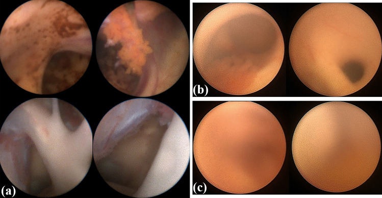
A retrospective analysis of all pediatric patients undergoing ventricular catheter placement using the ShuntScope from 01/2012 to 01/2022 in the author's department was performed. Demographic, clinical, and radiological data were evaluated. The visualization quality of the intraoperative endoscopy was stratified into the categories of excellent, medium, and poor and compared to the postoperative catheter tip placement. Follow-up evaluation included the surgical revision rate due to proximal catheter occlusion.
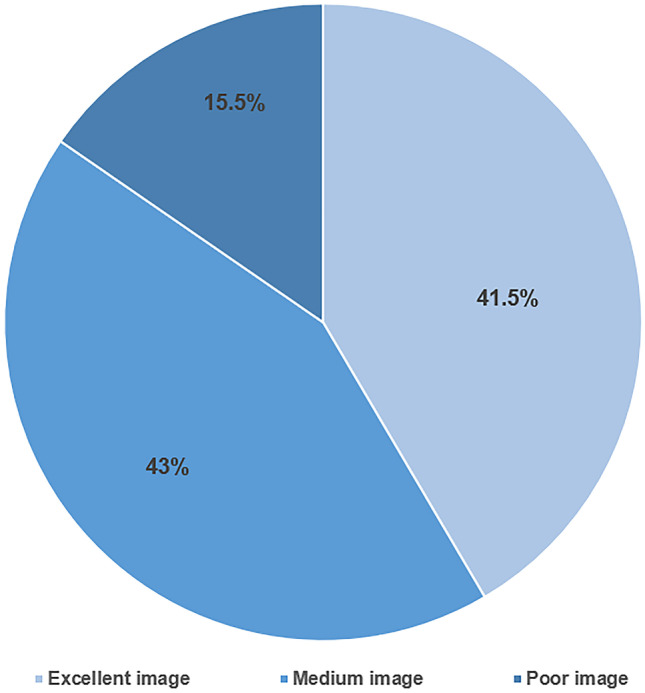
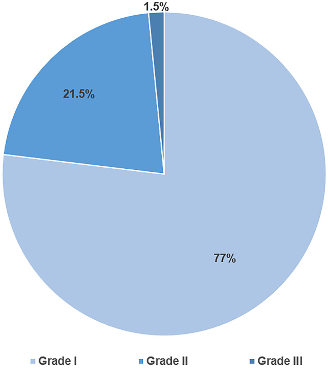
A total of 65 ShuntScope-assisted surgeries have been performed on 51 children. The mean age was 5.1 years. The most common underlying pathology was a tumor- or cyst-related hydrocephalus in 51%. Achieved image quality was excellent in 41.5%, medium in 43%, and poor in 15.5%. Ideal catheter placement was achieved in 77%. There were no intraoperative complications and no technique-related morbidity associated with the ShuntScope. The revision rate due to proximal occlusion was 4.61% during a mean follow-up period of 39.7 years. No statistical correlation between image grade and accuracy of catheter position was observed (p-value was 0.290).
The ShuntScope can be considered a valuable addition to standard surgical tools in treating pediatric hydrocephalus. Even suboptimal visualization contributes to high rates of correct catheter placement and, thereby, to a favorable clinical outcome.
由于解剖结构异常或需要进行经导水管支架放置,小儿脑积水患者的脑室导管植入可能成为一项极具挑战性的任务。ShuntScope 经腔内镜的发明旨在提高导管放置的成功率。本研究旨在评估 ShuntScope 在儿科人群中的图像质量和相关手术结果。
对作者所在科室 2012 年 1 月至 2022 年 1 月期间所有使用 ShuntScope 进行脑室导管放置的儿科患者进行回顾性分析。评估了人口统计学、临床和影像学数据。将术中内镜的可视化质量分为优秀、中等和差,并与术后导管尖端放置进行比较。随访评估包括因近端导管阻塞而进行的手术修正率。
共有 65 例 ShuntScope 辅助手术在 51 例儿童中进行。平均年龄为 5.1 岁。最常见的基础病理是肿瘤或囊肿相关脑积水,占 51%。获得的图像质量为优秀占 41.5%,中等占 43%,差占 15.5%。理想的导管放置率为 77%。术中无并发症,ShuntScope 无相关技术发病率。平均随访 39.7 年后近端阻塞的修正率为 4.61%。未观察到图像等级与导管位置准确性之间存在统计学相关性(p 值为 0.290)。
ShuntScope 可被视为治疗小儿脑积水标准手术工具的有价值的补充。即使是次优的可视化效果,也有助于实现高比例的正确导管放置,并由此获得良好的临床结果。