• 文献检索
  • 文档翻译
  • 深度研究
  • 学术资讯
  • Suppr Zotero 插件Zotero 插件
  • 邀请有礼
  • 套餐&价格
  • 历史记录
应用&插件
Suppr Zotero 插件Zotero 插件浏览器插件Mac 客户端Windows 客户端微信小程序
定价
高级版会员购买积分包购买API积分包
服务
文献检索文档翻译深度研究API 文档MCP 服务
关于我们
关于 Suppr公司介绍联系我们用户协议隐私条款
关注我们

Suppr 超能文献

核心技术专利:CN118964589B侵权必究
粤ICP备2023148730 号-1Suppr @ 2026

文献检索

告别复杂PubMed语法,用中文像聊天一样搜索,搜遍4000万医学文献。AI智能推荐,让科研检索更轻松。

立即免费搜索

文件翻译

保留排版,准确专业,支持PDF/Word/PPT等文件格式,支持 12+语言互译。

免费翻译文档

深度研究

AI帮你快速写综述,25分钟生成高质量综述,智能提取关键信息,辅助科研写作。

立即免费体验

胎盘炎症导致胚胎心脏发育异常。

Placental Inflammation Leads to Abnormal Embryonic Heart Development.

机构信息

William Harvey Research Institute, Queen Mary University of London, Charterhouse Square, UK (E.J.W., S.B., S.F., C.G., N.P.D., M.P., F.M.M.-B., S.N.).

European Bioinformatics Institute, Wellcome Genome Campus, Hinxton, Cambridge, UK (K.M.M.).

出版信息

Circulation. 2023 Mar 21;147(12):956-972. doi: 10.1161/CIRCULATIONAHA.122.061934. Epub 2022 Dec 9.

DOI:10.1161/CIRCULATIONAHA.122.061934
PMID:36484244
原文链接:https://pmc.ncbi.nlm.nih.gov/articles/PMC10022676/
Abstract

BACKGROUND

Placental heart development and embryonic heart development occur in parallel, and these organs have been proposed to exert reciprocal regulation during gestation. Poor placentation has been associated with congenital heart disease, an important cause of infant mortality. However, the mechanisms by which altered placental development can lead to congenital heart disease remain unresolved.

METHODS

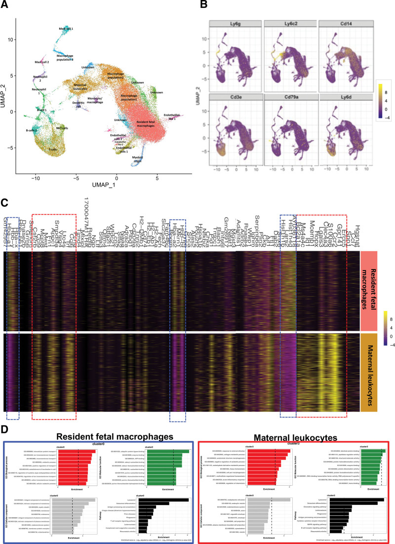
In this study, we use an in vivo neutrophil-driven placental inflammation model through antibody depletion of maternal circulating neutrophils at key stages during time-mated murine pregnancy: embryonic days 4.5 and 7.5. Pregnant mice were culled at embryonic day 14.5 to assess placental and embryonic heart development. A combination of flow cytometry, histology, and bulk RNA sequencing was used to assess placental immune cell composition and tissue architecture. We also used flow cytometry and single-cell sequencing to assess embryonic cardiac immune cells at embryonic day 14.5 and histology and gene analyses to investigate embryonic heart structure and development. In some cases, offspring were culled at postnatal days 5 and 28 to assess any postnatal cardiac changes in immune cells, structure, and cardiac function, as measured by echocardiography.

RESULTS

In the present study, we show that neutrophil-driven placental inflammation leads to inadequate placental development and loss of barrier function. Consequently, placental inflammatory monocytes of maternal origin become capable of migration to the embryonic heart and alter the normal composition of resident cardiac macrophages and cardiac tissue structure. This cardiac impairment continues into postnatal life, hindering normal tissue architecture and function. Last, we show that tempering placental inflammation can prevent this fetal cardiac defect and is sufficient to promote normal cardiac function in postnatal life.

CONCLUSIONS

Taken together, these observations provide a mechanistic paradigm whereby neutrophil-driven inflammation in pregnancy can preclude normal embryonic heart development as a direct consequence of poor placental development, which has major implications on cardiac function into adult life.

摘要

背景

胎盘心脏发育和胚胎心脏发育是平行发生的,这两个器官在妊娠期间被认为存在相互调节作用。不良的胎盘形成与先天性心脏病有关,后者是婴儿死亡的一个重要原因。然而,改变胎盘发育如何导致先天性心脏病的机制仍未解决。

方法

在这项研究中,我们使用了一种体内中性粒细胞驱动的胎盘炎症模型,通过在时间匹配的小鼠妊娠的关键阶段耗尽母体循环中性粒细胞来实现:胚胎第 4.5 天和第 7.5 天。在胚胎第 14.5 天处死怀孕小鼠以评估胎盘和胚胎心脏发育。我们使用流式细胞术、组织学和批量 RNA 测序来评估胎盘免疫细胞组成和组织结构。我们还使用流式细胞术和单细胞测序在胚胎第 14.5 天评估胚胎心脏免疫细胞,使用组织学和基因分析来研究胚胎心脏结构和发育。在某些情况下,在产后第 5 天和第 28 天处死后代,以评估免疫细胞、结构和心脏功能的任何产后变化,通过超声心动图进行测量。

结果

在本研究中,我们表明中性粒细胞驱动的胎盘炎症导致胎盘发育不足和屏障功能丧失。因此,母体来源的胎盘炎症单核细胞能够迁移到胚胎心脏,并改变常驻心脏巨噬细胞的正常组成和心脏组织结构。这种心脏损伤持续到产后,阻碍了正常的组织结构和功能。最后,我们表明,减轻胎盘炎症可以预防这种胎儿心脏缺陷,并足以促进产后的正常心脏功能。

结论

综上所述,这些观察结果提供了一个机制范例,即在妊娠期间,中性粒细胞驱动的炎症可以阻止正常的胚胎心脏发育,这是由于不良的胎盘发育直接导致的,这对成年后的心脏功能有重大影响。

https://cdn.ncbi.nlm.nih.gov/pmc/blobs/82f8/10022676/3f9da4568539/cir-147-956-g008.jpg
https://cdn.ncbi.nlm.nih.gov/pmc/blobs/82f8/10022676/339a2747c301/cir-147-956-g001.jpg
https://cdn.ncbi.nlm.nih.gov/pmc/blobs/82f8/10022676/a6161f0f06c0/cir-147-956-g002.jpg
https://cdn.ncbi.nlm.nih.gov/pmc/blobs/82f8/10022676/80de2aecc5b2/cir-147-956-g004.jpg
https://cdn.ncbi.nlm.nih.gov/pmc/blobs/82f8/10022676/3e4a140d2cfd/cir-147-956-g005.jpg
https://cdn.ncbi.nlm.nih.gov/pmc/blobs/82f8/10022676/d41cddeabcce/cir-147-956-g006.jpg
https://cdn.ncbi.nlm.nih.gov/pmc/blobs/82f8/10022676/56131f62d496/cir-147-956-g007.jpg
https://cdn.ncbi.nlm.nih.gov/pmc/blobs/82f8/10022676/3f9da4568539/cir-147-956-g008.jpg

相似文献

1
Placental Inflammation Leads to Abnormal Embryonic Heart Development.胎盘炎症导致胚胎心脏发育异常。
Circulation. 2023 Mar 21;147(12):956-972. doi: 10.1161/CIRCULATIONAHA.122.061934. Epub 2022 Dec 9.
2
Placental delayed villous maturation is associated with fetal congenital heart disease.胎盘绒毛成熟延迟与胎儿先天性心脏病有关。
Am J Obstet Gynecol. 2023 Feb;228(2):231.e1-231.e11. doi: 10.1016/j.ajog.2022.08.013. Epub 2022 Aug 17.
3
Heart defects and embryonic lethality in Asb2 knock out mice correlate with placental defects.Asb2基因敲除小鼠的心脏缺陷和胚胎致死率与胎盘缺陷相关。
Cells Dev. 2021 Mar;165:203663. doi: 10.1016/j.cdev.2021.203663. Epub 2021 Jan 27.
4
High-fat diet intake modulates maternal intestinal adaptations to pregnancy and results in placental hypoxia, as well as altered fetal gut barrier proteins and immune markers.高脂肪饮食摄入会调节母体肠道对妊娠的适应性,导致胎盘缺氧,以及胎儿肠道屏障蛋白和免疫标志物的改变。
J Physiol. 2019 Jun;597(12):3029-3051. doi: 10.1113/JP277353. Epub 2019 May 13.
5
Novel insights from fetal and placental phenotyping in 3 mouse models of Down syndrome.在 3 种唐氏综合征小鼠模型中对胎儿和胎盘表型的新见解。
Am J Obstet Gynecol. 2021 Sep;225(3):296.e1-296.e13. doi: 10.1016/j.ajog.2021.03.019. Epub 2021 Mar 22.
6
Placenta morphology and biomarkers in pregnancies with congenital heart disease - A systematic review.先天性心脏病妊娠的胎盘形态和生物标志物——系统评价。
Placenta. 2021 Sep 1;112:189-196. doi: 10.1016/j.placenta.2021.07.297. Epub 2021 Aug 8.
7
Advanced magnetic resonance imaging detects altered placental development in pregnancies affected by congenital heart disease.高级磁共振成像检测到先天性心脏病胎儿的胎盘发育改变。
Sci Rep. 2024 May 29;14(1):12357. doi: 10.1038/s41598-024-63087-8.
8
A mouse model of antepartum stillbirth.产前死产的小鼠模型。
Am J Obstet Gynecol. 2017 Oct;217(4):443.e1-443.e11. doi: 10.1016/j.ajog.2017.06.009. Epub 2017 Jun 12.
9
Conditional Mutation of Hand1 in the Mouse Placenta Disrupts Placental Vascular Development Resulting in Fetal Loss in Both Early and Late Pregnancy.条件性突变小鼠胎盘 Hand1 导致胎盘血管发育异常,导致早、晚期妊娠胎儿丢失。
Int J Mol Sci. 2021 Sep 2;22(17):9532. doi: 10.3390/ijms22179532.
10
Placental vascular malperfusion lesions in fetal congenital heart disease.胎儿先天性心脏病的胎盘血管灌注不良病变。
Am J Obstet Gynecol. 2022 Oct;227(4):620.e1-620.e8. doi: 10.1016/j.ajog.2022.05.038. Epub 2022 May 21.

引用本文的文献

1
Neurodevelopment in congenital heart disease: a review of antenatal mechanisms and therapeutic potentials.先天性心脏病中的神经发育:产前机制及治疗潜力综述
Pediatr Res. 2025 Sep 11. doi: 10.1038/s41390-025-04360-y.
2
Is maternal SARS-CoV-2 infection in the first trimester associated with congenital heart defects?孕早期母体感染新型冠状病毒肺炎是否与先天性心脏缺陷有关?
Front Pediatr. 2025 Aug 18;13:1643423. doi: 10.3389/fped.2025.1643423. eCollection 2025.
3
Prenatal Bisphenol A Exposure Impairs Fetal Heart Development: Molecular and Structural Alterations with Sex-Specific Differences.
产前双酚A暴露损害胎儿心脏发育:具有性别特异性差异的分子和结构改变
Antioxidants (Basel). 2025 Jul 14;14(7):863. doi: 10.3390/antiox14070863.
4
Placental Inflammation in Preterm Premature Rupture of Membranes and Risk of Neurodevelopmental Disorders.胎膜早破时的胎盘炎症与神经发育障碍风险
Cells. 2025 Jun 24;14(13):965. doi: 10.3390/cells14130965.
5
Placental histology, perioperative brain development, and neurodevelopmental outcome at 1 year of age in patients undergoing neonatal cardiac surgery-is there an association?新生儿心脏手术患者的胎盘组织学、围手术期脑发育及1岁时的神经发育结局——它们之间有关联吗?
Front Cardiovasc Med. 2025 May 30;12:1556289. doi: 10.3389/fcvm.2025.1556289. eCollection 2025.
6
Feto-placental blood vessel development.胎儿-胎盘血管发育
Development. 2025 Jun 1;152(11). doi: 10.1242/dev.204838. Epub 2025 Jun 2.
7
Genetic and Environmental Contributors To Congenital Heart Disease.先天性心脏病的遗传和环境因素
Curr Treat Options Cardiovasc Med. 2025;27(1):36. doi: 10.1007/s11936-025-01091-5. Epub 2025 May 26.
8
Pregnancy Complications in Fetal Congenital Heart Disease: A Result of Common Early Developmental Pathways Rather Than Fetal Hemodynamics.胎儿先天性心脏病中的妊娠并发症:常见早期发育途径的结果而非胎儿血流动力学的结果
Prenat Diagn. 2025 Aug;45(9):1122-1129. doi: 10.1002/pd.6819. Epub 2025 May 26.
9
Placental and cord serum inflammatory cytokines and children's domain-specific neurodevelopment at 18 months: effect modification by maternal vitamin D status.胎盘和脐带血清炎症细胞因子与18个月大儿童特定领域的神经发育:母体维生素D状态的效应修正
BMC Med. 2025 Apr 30;23(1):252. doi: 10.1186/s12916-025-04096-w.
10
Utero-placental (vascular) development throughout pregnancy in women with polycystic ovary syndrome: the Rotterdam Periconceptional Cohort.多囊卵巢综合征女性孕期子宫胎盘(血管)发育:鹿特丹围孕期队列研究
F S Rep. 2024 Dec 19;6(1):79-89. doi: 10.1016/j.xfre.2024.12.002. eCollection 2025 Mar.