Obray J Daniel, Landin Justine D, Vaughan Dylan T, Scofield Michael D, Chandler L Judson
Department of Neuroscience, Medical University of South Carolina, 30 Courtenay Drive, Charleston SC 29425, USA.
Department of Anesthesiology, Medical University of South Carolina, Charleston SC, USA.
Addict Neurosci. 2022 Dec;4. doi: 10.1016/j.addicn.2022.100044. Epub 2022 Nov 17.
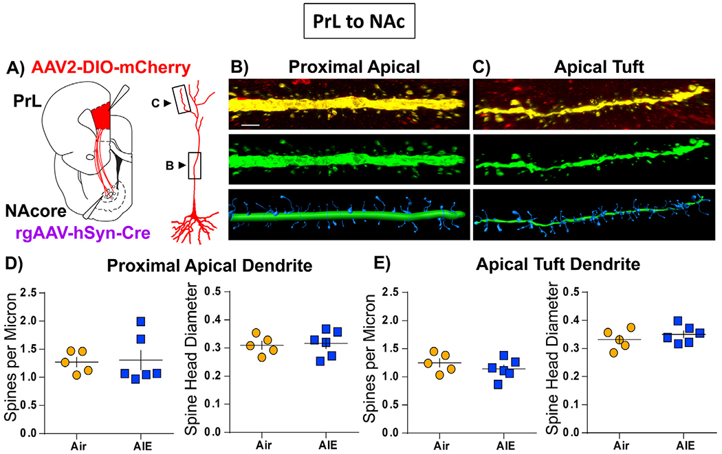
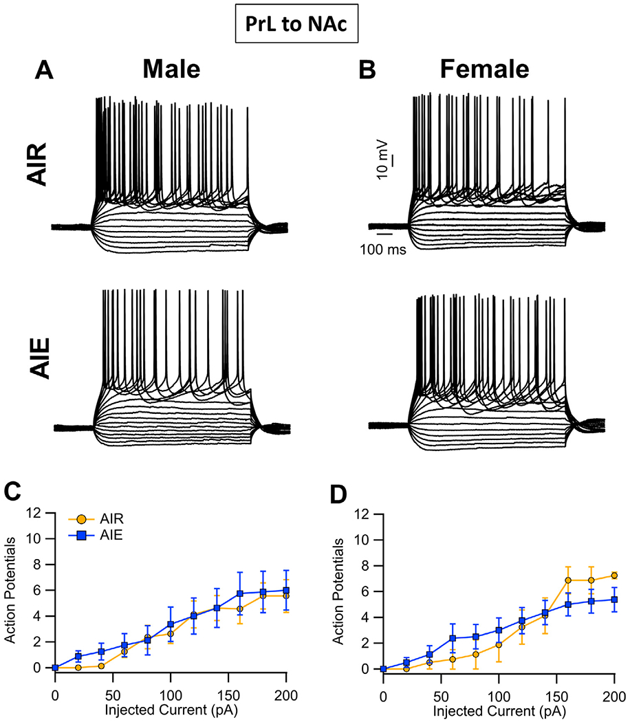
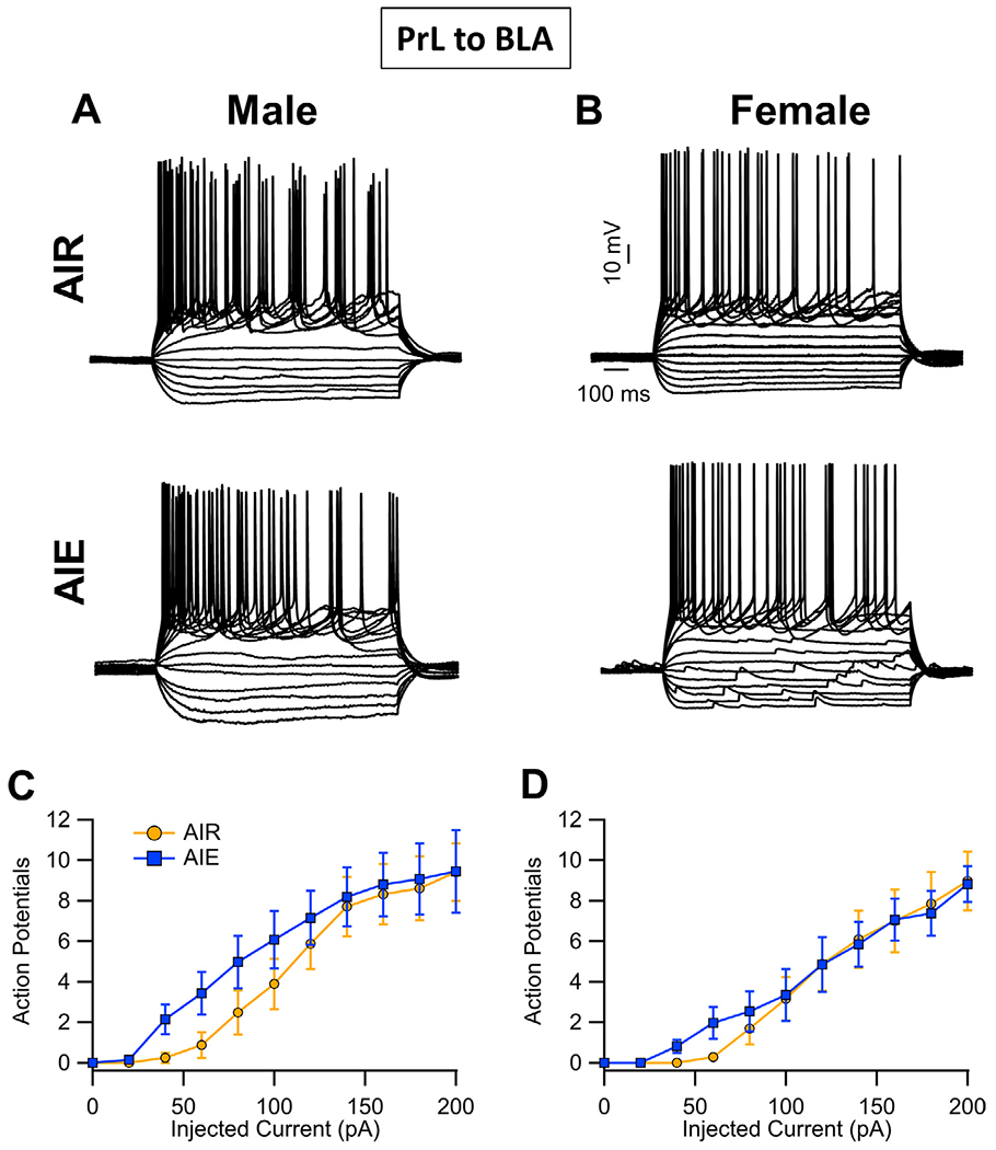
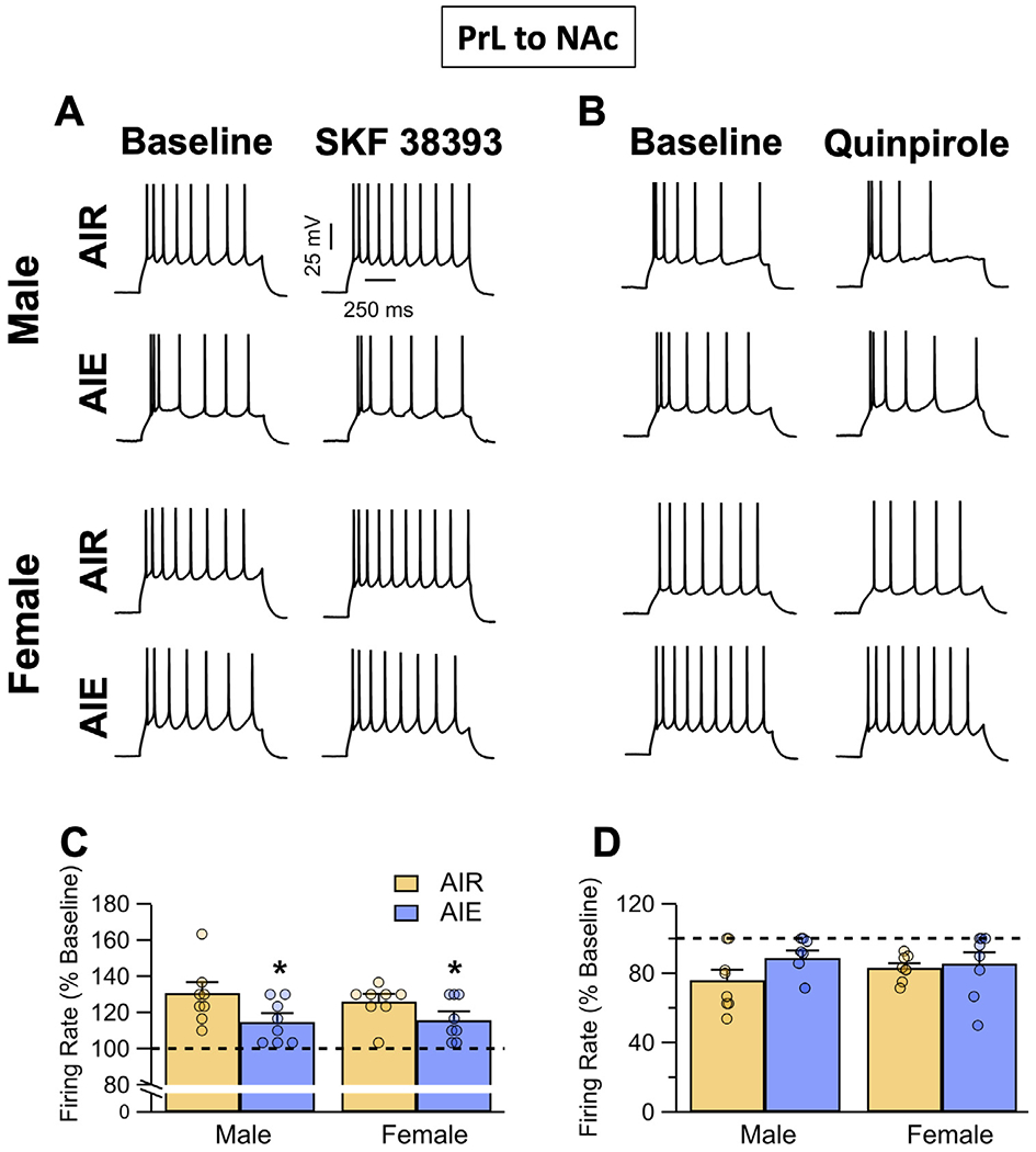
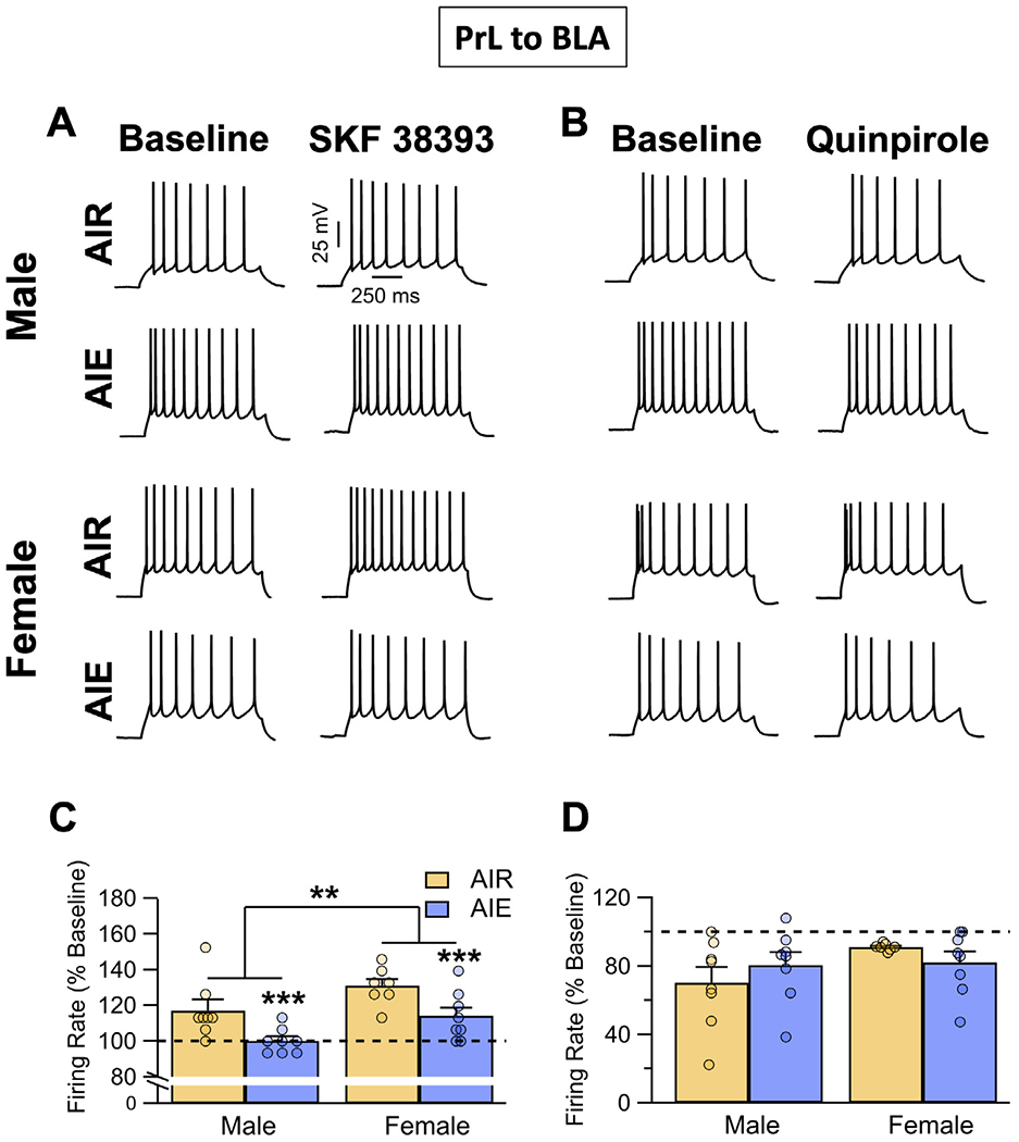
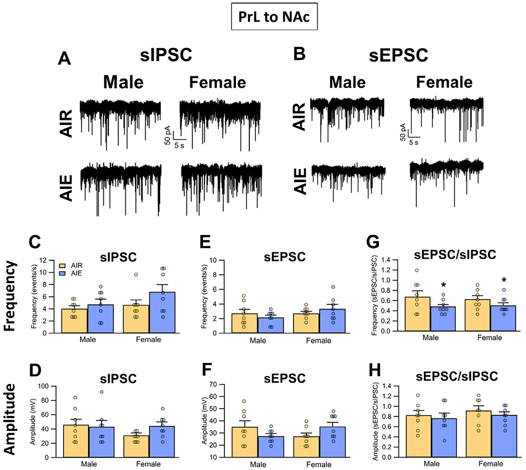
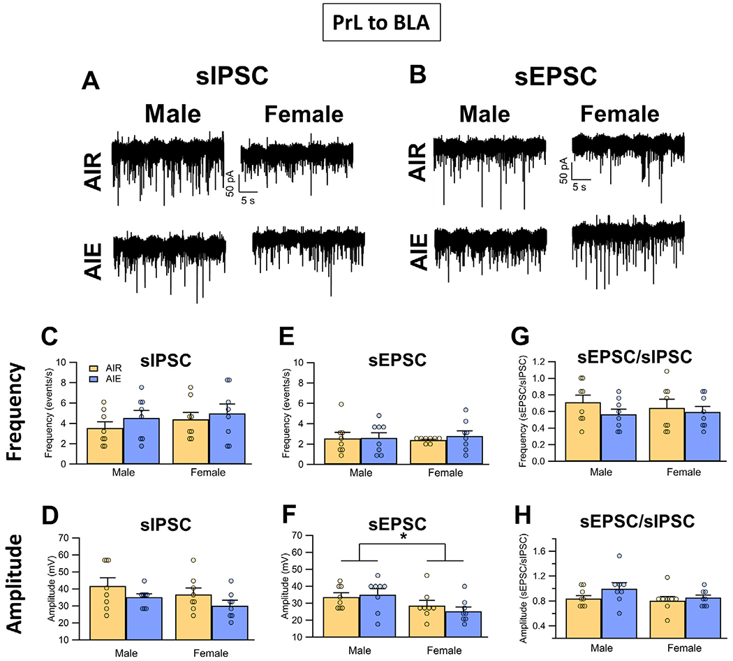
Binge drinking during adolescence is highly prevalent despite increasing evidence of its long-term impact on behaviors associated with modulation of behavioral flexibility by the medial prefrontal cortex (mPFC). In the present study, male and female rats underwent adolescent intermittent ethanol (AIE) exposure by vapor inhalation. After aging to adulthood, retrograde bead labelling and viral tagging were used to identify populations of neurons in the prelimbic region (PrL) of the mPFC that project to specific subcortical targets. Electrophysiological recording from bead-labelled neurons in PrL slices revealed that AIE did not alter the intrinsic excitability of PrL neurons that projected to either the NAc or the BLA. Similarly, recordings of spontaneous inhibitory and excitatory post-synaptic currents revealed no AIE-induced changes in synaptic drive onto either population of projection neurons. In contrast, AIE exposure was associated with a loss of dopamine receptor 1 (D1), but no change in dopamine receptor 2 (D2), modulation of evoked firing of both populations of projection neurons. Lastly, confocal imaging of proximal and apical dendritic tufts of viral-labelled PrL neurons that projected to the nucleus accumbens (NAc) revealed AIE did not alter the density of dendritic spines. Together, these observations provide evidence that AIE exposure results in disruption of D1 receptor modulation of PrL inputs to at least two major subcortical target regions that have been implicated in AIE-induced long-term changes in behavioral control.
尽管越来越多的证据表明青少年期狂饮对与内侧前额叶皮质(mPFC)调节行为灵活性相关的行为有长期影响,但青少年期狂饮现象仍然非常普遍。在本研究中,雄性和雌性大鼠通过蒸汽吸入接受青少年间歇性乙醇(AIE)暴露。成年后,采用逆行微珠标记和病毒标记来识别mPFC前边缘区(PrL)中投射到特定皮质下靶点的神经元群体。对PrL切片中微珠标记神经元的电生理记录显示,AIE并未改变投射到伏隔核(NAc)或杏仁核基底外侧核(BLA)的PrL神经元固有兴奋性。同样,对自发抑制性和兴奋性突触后电流的记录显示,AIE并未引起投射神经元群体突触驱动的变化。相比之下,AIE暴露与多巴胺受体1(D1)的丧失有关,但多巴胺受体2(D2)没有变化,这两种投射神经元群体的诱发放电调节也没有变化。最后,对投射到伏隔核(NAc)的病毒标记PrL神经元近端和顶端树突棘的共聚焦成像显示,AIE并未改变树突棘密度。总之,这些观察结果表明,AIE暴露会导致PrL输入到至少两个主要皮质下靶区域的D1受体调节受到破坏,而这些区域与AIE诱导的行为控制长期变化有关。