Moreira Maia Lucas, Maria Toubes Kênia, Moreira Júnior Gil, Quadros Tonelli Stephanie, de Carvalho Machado Vinicius, Ferreira Silveira Frank, Nunes Eduardo
Department of Operative Dentistry, School of Dentistry, Federal University of Minas Gerais, Belo Horizonte, Minas Gerais, Brazil.
Department of Dentistry, Pontifícial Catholic University of Minas Gerais, Belo Horizonte, Minas Gerais, Brazil.
Iran Endod J. 2020 Spring;15(2):111-116. doi: 10.22037/iej.v15i2.27183.
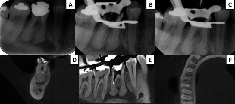
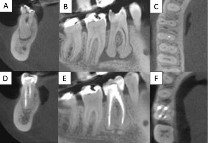
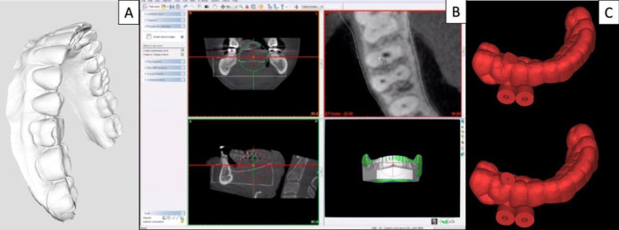
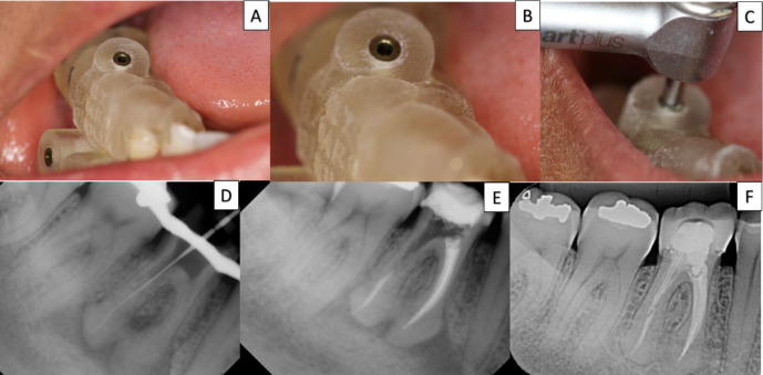
This case report describes the use of the guided endodontics for a non-surgical endodontic retreatment of the mandibular molar. A 38-year-old female reported apical swelling and localized pain on the tooth #30, exacerbated when chewing hard food. Periapical radiographic examination showed pulp canal obliteration in the apical third associated with extensive radiolucent area. Cone-beam computed tomography (CBCT) images were requested to support the diagnosis and enable preparation of a surgical guide, used to direct access to the canals that were obliterated and incompletely filled. The follow-up at 24 months radiographically showed completely healed apical area in the involved tooth. This non-surgical technique demonstrated efficacy in case resolution.
本病例报告描述了使用引导式牙髓治疗术对下颌磨牙进行非手术牙髓再治疗的情况。一名38岁女性报告称30号牙出现根尖肿胀和局部疼痛,咀嚼硬物时疼痛加剧。根尖X线片检查显示根尖三分之一处牙髓根管闭塞,并伴有广泛的透射区。请求进行锥形束计算机断层扫描(CBCT)成像以辅助诊断,并制作手术导板,用于引导进入闭塞和未完全充填的根管。术后24个月的X线片随访显示,患牙根尖区完全愈合。这种非手术技术在病例治疗中显示出了疗效。