Kato Ryohei, Kato Takahiro, Narita Yuki, Sasaki Sho, Takayama Kanako, Murakami Masao
Department of Radiation Physics and Technology, Southern Tohoku Proton Therapy Center, Fukushima, Japan.
Department of Radiological Sciences, School of Health Sciences, Fukushima Medical University, Fukushima, Japan.
Adv Radiat Oncol. 2022 Dec 25;8(4):101153. doi: 10.1016/j.adro.2022.101153. eCollection 2023 Jul-Aug.
To identify the induced radionuclides produced from dental metals in proton beam therapy and investigate the accuracy of the Monte Carlo (MC) simulation by comparing the measured radioactivity.
Two dental metals of pure titanium and gold-silver-palladium alloy, commonly used in Japan, were used in this study. The dental metal placed at the center of Spread-out Bragg Peak was irradiated by 150-MeV passive scattering proton beam. The gamma rays emitted from the activated dental metals were measured using a high purity germanium (HPGe) detector. The induced radionuclides were identified from the measured gamma-ray energies. Furthermore, the Particle and Heavy Ion Transport code System v.3.24 and DCHAIN were used for the MC simulation. The measured radionuclides and their radioactivity were compared with the simulation results.
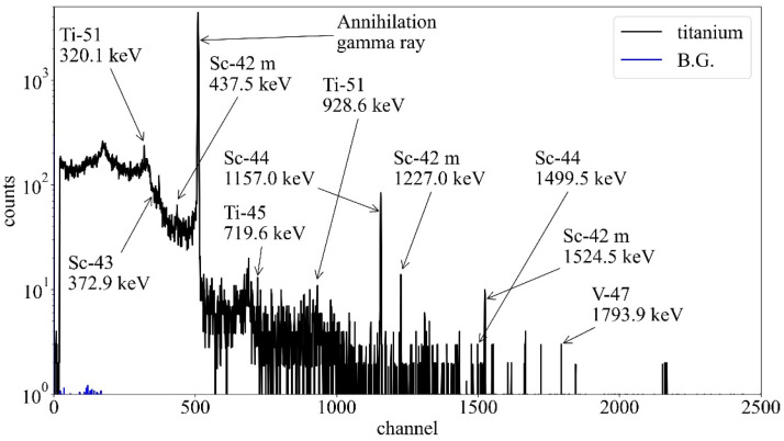
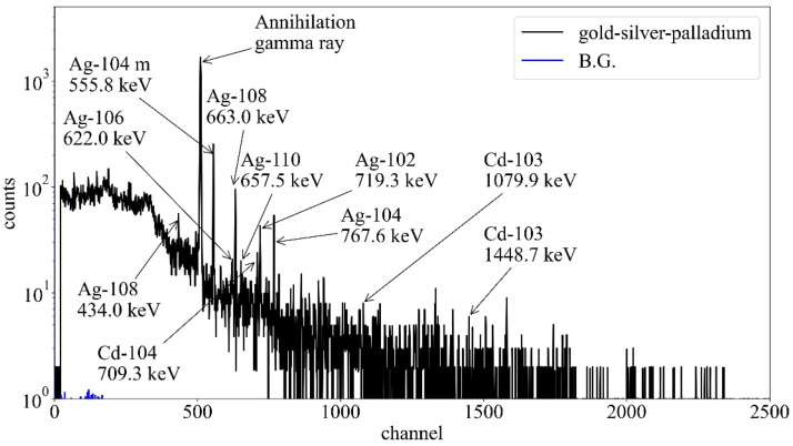
In the MC simulation for the activated titanium, vanadium-47, with a half-life of 32.6 minutes had the strongest radioactivity among the induced radionuclides. The energy peaks of gamma rays emitted from titanium-51, scandium-43, scandium-44, and annihilation gamma rays were observed for the activated titanium in the HPGe detector. In the MC simulation for the activated gold-silver-palladium alloy, silver-108, with a half-life of 2.4 minutes had the strongest radioactivity. The energy peaks of gamma rays emitted from silver-104, silver-104 m, silver-108, and annihilation gamma rays were observed for the activated gold-silver-palladium alloy in the HPGe detector. Furthermore, the induced radionuclides and their radioactivity in the MC simulation were consistent with the measurement results for both dental metals, except for a few radionuclides.
We identify the induced radionuclides produced from 2 dental metals and compared their radioactivity between the measurements and the MC simulation. Although the identification of the induced radionuclides using the MC simulation remains uncertain, the MC simulation can be clinically effective for pre-estimating the induced radionuclides in proton beam therapy.
识别质子束治疗中牙科金属产生的感生放射性核素,并通过比较测量的放射性来研究蒙特卡罗(MC)模拟的准确性。
本研究使用了日本常用的两种牙科金属,纯钛和金银钯合金。将牙科金属置于扩展布拉格峰中心,用150 MeV被动散射质子束进行照射。使用高纯锗(HPGe)探测器测量活化牙科金属发射的伽马射线。从测量的伽马射线能量中识别感生放射性核素。此外,使用粒子和重离子传输代码系统v.3.24和DCHAIN进行MC模拟。将测量的放射性核素及其放射性与模拟结果进行比较。
在活化钛的MC模拟中,半衰期为32.6分钟的钒-47在感生放射性核素中具有最强的放射性。在HPGe探测器中观察到活化钛发射的钛-51、钪-43、钪-44的伽马射线能量峰以及湮没伽马射线。在活化金银钯合金的MC模拟中,半衰期为2.4分钟的银-108具有最强的放射性。在HPGe探测器中观察到活化金银钯合金发射的银-104、银-104m、银-108的伽马射线能量峰以及湮没伽马射线。此外,除了少数放射性核素外,两种牙科金属的MC模拟中的感生放射性核素及其放射性与测量结果一致。
我们识别了两种牙科金属产生的感生放射性核素,并比较了测量值与MC模拟之间的放射性。虽然使用MC模拟识别感生放射性核素仍不确定,但MC模拟在质子束治疗中对预估算感生放射性核素可能具有临床有效性。