Department of Biology, University of Victoria, Victoria, BC, Canada.
University of Illinois College of Medicine, Chicago, IL, United States.
Front Immunol. 2023 Feb 6;14:1050594. doi: 10.3389/fimmu.2023.1050594. eCollection 2023.
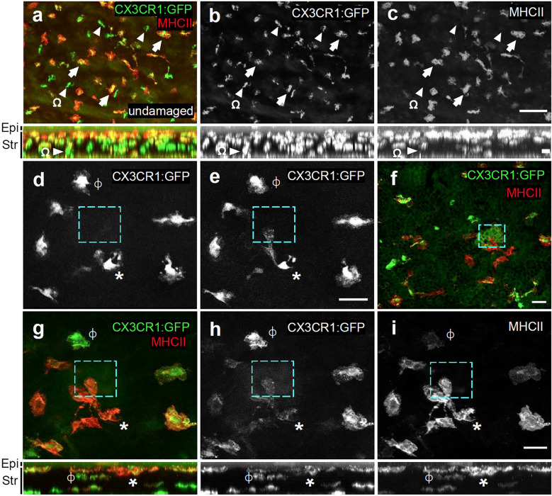
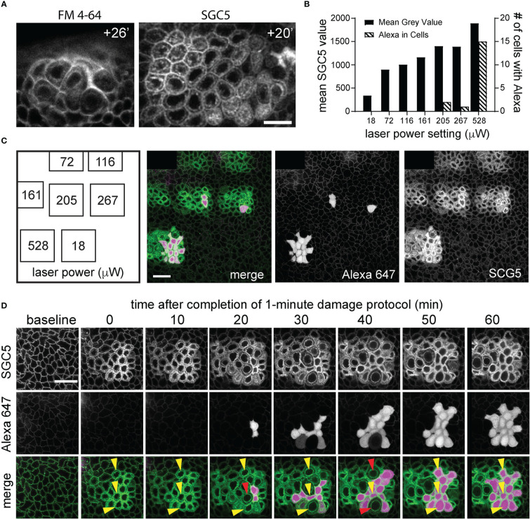
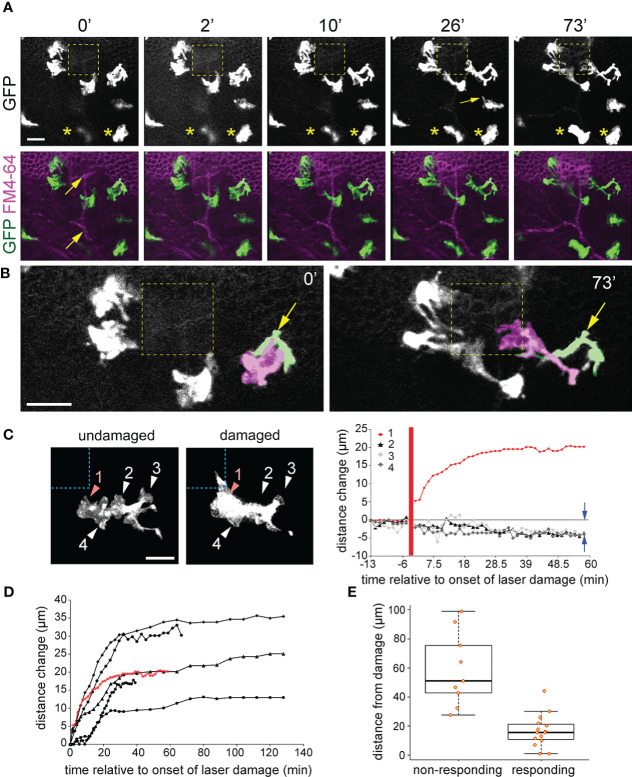
The corneal epithelium is continuously subjected to external stimuli that results in varying degrees of cellular damage. The use of live-cell imaging approaches has facilitated understanding of the cellular and molecular mechanisms underlying the corneal epithelial wound healing process. Here, we describe a live, , whole-eye approach using laser scanning confocal microscopy to simultaneously induce and visualize short-term cellular responses following microdamage to the corneal epithelium. Live-cell imaging of corneal cell layers was enabled using the lipophilic fluorescent dyes, SGC5 or FM4-64, which, when injected into the anterior chamber of enucleated eyes, readily penetrated and labelled cell membranes. Necrotic microdamage to a defined region (30 μm x 30 μm) through the central plane of the corneal basal epithelium was induced by continuously scanning for at least one minute using high laser power and was dependent on the presence of lipophilic fluorescent dye. This whole-mount live-cell imaging and microdamage approach was used to examine the behavior of -expressing resident corneal stromal macrophages (RCSMs). In undamaged corneas, RCSMs remained stationary, but exhibited a constant extension and retraction of short (~5 μm) semicircular, pseudopodia-like processes reminiscent of what has previously been reported in corneal dendritic cells. Within minutes of microdamage, nearby anterior RCSMs became highly polarized and extended projections towards the damaged region. The extension of the processes plateaued after about 30 minutes and remained stable over the course of 2-3 hours of imaging. Retrospective immunolabeling showed that these responding RCSMs were MHC class II+. This study adds to existing knowledge of immune cell behavior in response to corneal damage and introduces a simple corneal epithelial microdamage and wound healing paradigm.
角膜上皮不断受到外部刺激,导致细胞损伤程度不同。使用活细胞成像方法有助于理解角膜上皮伤口愈合过程中的细胞和分子机制。在这里,我们描述了一种使用激光扫描共聚焦显微镜的活体、全眼方法,该方法可同时诱导和可视化角膜上皮微损伤后短期的细胞反应。使用亲脂性荧光染料 SGC5 或 FM4-64 对角膜细胞层进行活细胞成像,这些染料在注入眼前房后很容易穿透并标记细胞膜。通过使用高激光功率连续扫描至少一分钟,在角膜基底层的中央平面上对一个确定的区域(30μm×30μm)进行坏死性微损伤,这种微损伤依赖于亲脂性荧光染料的存在。使用这种全层活细胞成像和微损伤方法来研究表达的固有角膜基质巨噬细胞(RCSM)的行为。在未受损的角膜中,RCSM 保持静止,但表现出短(约 5μm)半圆形伪足样突起的恒定延伸和回缩,这与以前在角膜树突状细胞中报道的相似。在微损伤后的几分钟内,附近的前 RCSM 变得高度极化,并向受损区域延伸突起。突起的延伸在大约 30 分钟后达到平台期,并在 2-3 小时的成像过程中保持稳定。回顾性免疫标记显示,这些反应性 RCSM 是 MHC Ⅱ类阳性的。本研究增加了对免疫细胞在角膜损伤反应中的行为的现有认识,并引入了一种简单的角膜上皮微损伤和伤口愈合模型。