• 文献检索
  • 文档翻译
  • 深度研究
  • 学术资讯
  • Suppr Zotero 插件Zotero 插件
  • 邀请有礼
  • 套餐&价格
  • 历史记录
应用&插件
Suppr Zotero 插件Zotero 插件浏览器插件Mac 客户端Windows 客户端微信小程序
定价
高级版会员购买积分包购买API积分包
服务
文献检索文档翻译深度研究API 文档MCP 服务
关于我们
关于 Suppr公司介绍联系我们用户协议隐私条款
关注我们

Suppr 超能文献

核心技术专利:CN118964589B侵权必究
粤ICP备2023148730 号-1Suppr @ 2026

文献检索

告别复杂PubMed语法,用中文像聊天一样搜索,搜遍4000万医学文献。AI智能推荐,让科研检索更轻松。

立即免费搜索

文件翻译

保留排版,准确专业,支持PDF/Word/PPT等文件格式,支持 12+语言互译。

免费翻译文档

深度研究

AI帮你快速写综述,25分钟生成高质量综述,智能提取关键信息,辅助科研写作。

立即免费体验

脂肪坏死:会诊医生的难题。

Fat necrosis: A consultant's conundrum.

作者信息

Majithia Jinita, Haria Purvi, Popat Palak, Katdare Aparna, Chouhan Sonal, Gala Kunal Bharat, Kulkarni Suyash, Thakur Meenakshi

机构信息

Radiology Department, Tata Memorial Hospital, Mumbai, India.

出版信息

Front Oncol. 2023 Feb 16;12:926396. doi: 10.3389/fonc.2022.926396. eCollection 2022.

DOI:10.3389/fonc.2022.926396
PMID:36873302
原文链接:https://pmc.ncbi.nlm.nih.gov/articles/PMC9978799/
Abstract

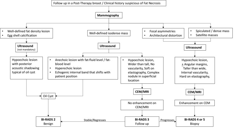
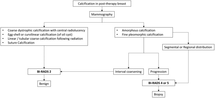
Fat necrosis of the breast is a benign non-suppurative inflammation of the adipose tissue and often mimics breast cancers, posing a diagnostic challenge for the clinician and radiologist. It has a myriad of appearances on different imaging techniques, ranging from the pathognomic oil cyst and benign dystrophic calcifications to indeterminate focal asymmetries, architectural distortions, and masses. A combination of different modalities can assist a radiologist in reaching a logical conclusion to avoid unnecessary interventions. The aim of this review article was to provide a comprehensive literature on the various imaging appearances of fat necrosis in the breast. Although a purely benign entity, the imaging appearances on mammography, contrast-enhanced mammography, ultrasound, and magnetic resonance imaging can be quite misleading, especially in post-therapy breasts. The purpose is to provide a comprehensive and all-inclusive review on fat necrosis with a proposed algorithm allowing a systematic approach to diagnosis.

摘要

乳腺脂肪坏死是脂肪组织的一种良性非化脓性炎症,常酷似乳腺癌,给临床医生和放射科医生带来诊断挑战。在不同的成像技术上,它有各种各样的表现,从典型的油囊肿和良性营养不良性钙化到不确定的局灶性不对称、结构扭曲和肿块。不同检查方式的联合应用有助于放射科医生得出合理结论,避免不必要的干预。这篇综述文章的目的是提供关于乳腺脂肪坏死各种成像表现的全面文献。尽管它是一种纯粹的良性病变,但乳腺钼靶、对比增强乳腺钼靶、超声和磁共振成像上的表现可能会极具误导性,尤其是在接受过治疗的乳腺中。目的是对脂肪坏死进行全面且涵盖所有方面的综述,并提出一种算法,以便采用系统的方法进行诊断。

https://cdn.ncbi.nlm.nih.gov/pmc/blobs/d713/9978799/afce9f7cd58e/fonc-12-926396-g002.jpg
https://cdn.ncbi.nlm.nih.gov/pmc/blobs/d713/9978799/8dc2e163e15c/fonc-12-926396-g001.jpg
https://cdn.ncbi.nlm.nih.gov/pmc/blobs/d713/9978799/afce9f7cd58e/fonc-12-926396-g002.jpg
https://cdn.ncbi.nlm.nih.gov/pmc/blobs/d713/9978799/8dc2e163e15c/fonc-12-926396-g001.jpg
https://cdn.ncbi.nlm.nih.gov/pmc/blobs/d713/9978799/afce9f7cd58e/fonc-12-926396-g002.jpg

相似文献

1
Fat necrosis: A consultant's conundrum.脂肪坏死:会诊医生的难题。
Front Oncol. 2023 Feb 16;12:926396. doi: 10.3389/fonc.2022.926396. eCollection 2022.
2
A pictorial review: multimodality imaging of benign and suspicious features of fat necrosis in the breast.图文综述:乳腺脂肪坏死的良性及可疑特征的多模态成像
Br J Radiol. 2018 Dec;91(1092):20180213. doi: 10.1259/bjr.20180213. Epub 2018 Jul 31.
3
Imaging of fat-containing lesions of the breast: a pictorial essay.乳腺含脂肪病变的影像学:图文综述
J Clin Ultrasound. 2013 Sep;41(7):424-33. doi: 10.1002/jcu.22070. Epub 2013 Jul 8.
4
Fat necrosis of the breast: clinical, mammographic and sonographic features.乳腺脂肪坏死:临床、乳腺X线及超声特征
Eur J Radiol. 2001 Aug;39(2):92-9. doi: 10.1016/s0720-048x(00)00303-x.
5
Sonography of fat necrosis of the breast: correlation with mammography and MR imaging.乳腺脂肪坏死的超声检查:与乳腺X线摄影及磁共振成像的相关性
J Clin Ultrasound. 2013 Sep;41(7):415-23. doi: 10.1002/jcu.22061. Epub 2013 May 28.
6
Palpable Lumps after Mastectomy: Radiologic-Pathologic Review of Benign and Malignant Masses.乳房切除术后可触及的肿块:良性和恶性肿块的放射病理回顾。
Radiographics. 2021 Jul-Aug;41(4):967-989. doi: 10.1148/rg.2021200161. Epub 2021 May 14.
7
Radiologic findings in women after Autologous Fat Transfer (AFT) based breast reconstruction: A Systematic Review.基于自体脂肪移植(AFT)的乳房重建术后女性的放射学表现:一项系统评价。
JPRAS Open. 2024 Aug 20;42:113-132. doi: 10.1016/j.jpra.2024.08.002. eCollection 2024 Dec.
8
The mammographic spectrum of fat necrosis of the breast.乳腺脂肪坏死的乳腺X线表现谱。
Radiographics. 1995 Nov;15(6):1347-56. doi: 10.1148/radiographics.15.6.8577961.
9
Choice of imaging method in the work-up of non-calcified breast lesions identified on tomosynthesis screening.在数字化乳腺断层合成筛查中发现的非钙化性乳腺病变的影像学方法选择。
Eur J Radiol. 2020 Oct;131:109203. doi: 10.1016/j.ejrad.2020.109203. Epub 2020 Aug 5.
10
Diagnostic Evaluation of Recalled Noncalcified Lesions Using Ultrasound Alone Versus Ultrasound Plus Additional Mammographic Views: A Prospective Study.仅用超声与超声加额外乳腺 X 线视图对召回的非钙化病变进行诊断评估:一项前瞻性研究。
AJR Am J Roentgenol. 2022 Jun;218(6):977-987. doi: 10.2214/AJR.21.26700. Epub 2021 Dec 15.

引用本文的文献

1
The Role of Exclusive Autologous Lipotransfer in Non-Irradiated Breasts After Mastectomy.单纯自体脂肪移植在乳房切除术后未接受放疗的乳房中的作用。
J Clin Med. 2025 Jun 24;14(13):4468. doi: 10.3390/jcm14134468.
2
Melanisation in Salmonid Skeletal Muscle: A Review.鲑科鱼类骨骼肌的黑化:综述
J Fish Dis. 2025 Mar;48(3):e14063. doi: 10.1111/jfd.14063. Epub 2024 Dec 11.
3
Fat Necrosis in Single Perforator Deep Inferior Epigastric Artery and Superficial Epigastric Artery Perforator Free Flaps: A Prospective Randomized Study.

本文引用的文献

1
Appearance of fat necrosis on contrast-enhanced spectral mammography.对比增强光谱乳腺摄影中脂肪坏死的表现。
BMJ Case Rep. 2022 Mar 15;15(3):e246231. doi: 10.1136/bcr-2021-246231.
2
Time to development and imaging features of new calcifications in the treated breast after breast-conserving therapy.保乳治疗后新钙化灶的出现时间及影像学特征。
Breast J. 2021 Oct;27(10):761-767. doi: 10.1111/tbj.14277. Epub 2021 Aug 6.
3
Comparison of the sensitivity of mammography, ultrasound, magnetic resonance imaging and combinations of these imaging modalities for the detection of small (≤2 cm) breast cancer.
单穿支腹壁下深动脉和腹壁浅动脉穿支游离皮瓣中的脂肪坏死:一项前瞻性随机研究
Plast Reconstr Surg Glob Open. 2024 Sep 12;12(9):e6152. doi: 10.1097/GOX.0000000000006152. eCollection 2024 Sep.
4
Case report: Atypical lipomatous tumor of the thigh in a four-year-old girl.病例报告:一名4岁女孩大腿部的非典型脂肪瘤性肿瘤。
Front Oncol. 2024 Jul 23;14:1401861. doi: 10.3389/fonc.2024.1401861. eCollection 2024.
比较乳腺 X 线摄影、超声、磁共振成像以及这些影像学方法联合应用对小(≤2cm)乳腺癌的检测灵敏度。
Medicine (Baltimore). 2021 Jul 2;100(26):e26531. doi: 10.1097/MD.0000000000026531.
4
Risk Factors for Fat Necrosis After Stereotactic Partial Breast Irradiation for Early-Stage Breast Cancer in a Phase 1 Clinical Trial.立体定向部分乳房照射治疗早期乳腺癌 1 期临床试验中脂肪坏死的危险因素。
Int J Radiat Oncol Biol Phys. 2020 Nov 1;108(3):697-706. doi: 10.1016/j.ijrobp.2020.05.025. Epub 2020 May 26.
5
Fat necrosis in the Breast: A systematic review of clinical.乳房脂肪坏死:临床的系统评价。
Lipids Health Dis. 2019 Jun 11;18(1):139. doi: 10.1186/s12944-019-1078-4.
6
Contrast-enhanced spectral mammography in the evaluation of breast suspicious calcifications: diagnostic accuracy and impact on surgical management.对比增强光谱乳腺摄影术在评估乳腺可疑钙化中的应用:诊断准确性及对手术管理的影响
Acta Radiol. 2019 Sep;60(9):1110-1117. doi: 10.1177/0284185118822639. Epub 2019 Jan 24.
7
Breast imaging surveillance after curative treatment for primary non-metastasised breast cancer in non-high-risk women: a systematic review.非高危女性原发性非转移性乳腺癌根治性治疗后的乳腺影像监测:一项系统评价
Insights Imaging. 2018 Dec;9(6):961-970. doi: 10.1007/s13244-018-0667-5. Epub 2018 Nov 8.
8
Contrast-Enhanced Mammography: A Systematic Guide to Interpretation and Reporting.对比增强乳腺摄影:解读与报告的系统指南。
AJR Am J Roentgenol. 2019 Jan;212(1):222-231. doi: 10.2214/AJR.17.19265. Epub 2018 Nov 1.
9
Fat necrosis after breast-conserving oncoplastic surgery.保乳肿瘤整形术后脂肪坏死。
Breast Cancer. 2019 Jan;26(1):125-130. doi: 10.1007/s12282-018-0901-5. Epub 2018 Aug 27.
10
Optimizing Perforator Selection: A Multivariable Analysis of Predictors for Fat Necrosis and Abdominal Morbidity in DIEP Flap Breast Reconstruction.优化穿支皮瓣选择:DIEP 游离皮瓣乳房重建中脂肪坏死和腹部并发症的多变量预测因素分析。
Plast Reconstr Surg. 2018 Sep;142(3):583-592. doi: 10.1097/PRS.0000000000004631.