Yang Ruohan, Jia Lin, Lv Zheng, Cui Jiuwei
Cancer Center, the First Hospital of Jilin University, Changchun, China.
Front Surg. 2023 Feb 21;10:1025287. doi: 10.3389/fsurg.2023.1025287. eCollection 2023.
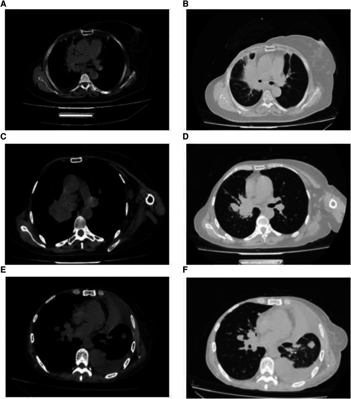
The lungs are a common metastatic organ in breast cancer, mainly due to blood metastasis. On imaging, most metastatic lesions show a peripheral round mass in the lung, occasionally with a hilar mass as the primary manifestation, showing burr and lobulation signs. This study aimed to investigate breast cancer patient's clinical characteristics and prognosis with two different metastatic sites in the lung.
We retrospectively analyzed patients admitted to the First Hospital of Jilin University between 2016 and 2021 diagnosed with breast cancer lung metastases. Forty breast cancer patients with hilar metastases (HM) and 40 patients with peripheral lung metastases (PLM) were matched 1:1 using a pairing method. To analyze the patient's prognosis, the clinical characteristics of patients with two different metastatic sites were compared using the chi-square test, Kaplan-Meier curve, and Cox proportional hazards model.
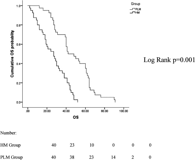
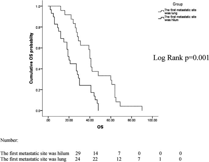
The median follow-up time was 38 months (2-91 months). The median age of patients with HM was 56 years (25-75 years), and that of patients with PLM was 59 years (44-82 years). The median overall survival (mOS) was 27 months in the HM group and 42 months in the PLM group ( = 0.001). The results of the Cox proportional hazards model showed that the histological grade (hazard ratio = 2.741, 95% confidence interval 1.442-5.208, = 0.002) was a prognostic factor in the HM group.
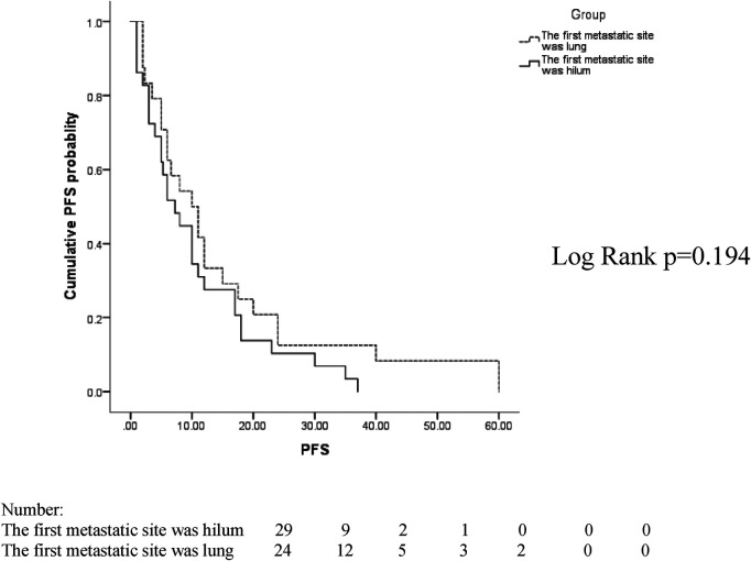
The number of young patients in the HM group was higher than that in the PLM group, with higher Ki-67 indexes and histological grades. Most patients had mediastinal lymph node metastasis, with shorter DFI and OS and poor prognosis.
肺是乳腺癌常见的转移器官,主要通过血行转移。在影像学上,大多数转移瘤表现为肺部外周圆形肿块,偶尔以肺门肿块为主要表现,可见毛刺征和分叶征。本研究旨在探讨乳腺癌患者肺内两种不同转移部位的临床特征及预后。
我们回顾性分析了2016年至2021年在吉林大学第一医院确诊为乳腺癌肺转移的患者。采用配对法将40例肺门转移(HM)的乳腺癌患者和40例肺外周转移(PLM)的患者进行1:1匹配。为分析患者的预后,采用卡方检验、Kaplan-Meier曲线和Cox比例风险模型比较了两种不同转移部位患者的临床特征。
中位随访时间为38个月(2 - 91个月)。HM组患者的中位年龄为56岁(25 - 75岁),PLM组患者的中位年龄为59岁(44 - 82岁)。HM组的中位总生存期(mOS)为27个月,PLM组为42个月(=0.001)。Cox比例风险模型结果显示,组织学分级(风险比=2.741,95%置信区间1.442 - 5.208,=0.002)是HM组的一个预后因素。
HM组年轻患者数量高于PLM组,Ki-67指数和组织学分级更高。大多数患者有纵隔淋巴结转移,无病生存期和总生存期较短,预后较差。