• 文献检索
  • 文档翻译
  • 深度研究
  • 学术资讯
  • Suppr Zotero 插件Zotero 插件
  • 邀请有礼
  • 套餐&价格
  • 历史记录
应用&插件
Suppr Zotero 插件Zotero 插件浏览器插件Mac 客户端Windows 客户端微信小程序
定价
高级版会员购买积分包购买API积分包
服务
文献检索文档翻译深度研究API 文档MCP 服务
关于我们
关于 Suppr公司介绍联系我们用户协议隐私条款
关注我们

Suppr 超能文献

核心技术专利:CN118964589B侵权必究
粤ICP备2023148730 号-1Suppr @ 2026

文献检索

告别复杂PubMed语法,用中文像聊天一样搜索,搜遍4000万医学文献。AI智能推荐,让科研检索更轻松。

立即免费搜索

文件翻译

保留排版,准确专业,支持PDF/Word/PPT等文件格式,支持 12+语言互译。

免费翻译文档

深度研究

AI帮你快速写综述,25分钟生成高质量综述,智能提取关键信息,辅助科研写作。

立即免费体验

爬行动物真皮骨的骨折修复:爬行动物能形成次生软骨吗?

Fracture repair of reptilian dermal bones: can reptiles form secondary cartilage?

作者信息

Irwin C R, Ferguson M W

机构信息

Department of Anatomy, Queen's University of Belfast.

出版信息

J Anat. 1986 Jun;146:53-64.

PMID:3693062
原文链接:https://pmc.ncbi.nlm.nih.gov/articles/PMC1166524/
Abstract

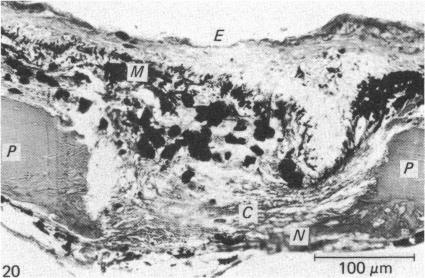
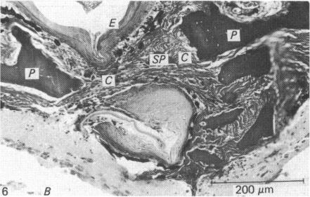
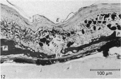
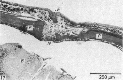
The fracture repair of reptilian dermal bones has not previously been reported. Moreover, repair of fractured dermal bones in birds and mammals involves secondary chondrogenesis whereas that of amphibians does not. Therefore an investigation into the repair of fractured reptilian dermal bones could reveal the stage during vertebrate evolution at which the process of secondary chondrogenesis appeared. Experimental incisions were made in the parietal bones of seventeen lizards (3 species) and 2 snakes (1 species). These resulted in a fracture environment of limited vascularity and increased movement--two known stimuli of secondary chondrogenesis in birds and mammals. Re-epithelialisation was rapid and dead bony fragments quickly sequestered. The blood blot was quickly organised into connective tissue, the dural periostea proliferated, osteoblasts differentiated and bony union was effected after 18 days. The width of the fracture gap was the principal variable affecting the chronology of fracture repair. Secondary cartilage was not detected in any specimen, of any species, at any stage of the fracture repair. It therefore appears that the progenitor cells on reptilian dermal bones are not capable of forming secondary cartilage and that this tissue arose comparatively late in vertebrate evolution.

摘要

此前尚未有关于爬行动物真皮骨骨折修复的报道。此外,鸟类和哺乳动物真皮骨骨折的修复涉及继发性软骨形成,而两栖动物则不然。因此,对爬行动物真皮骨骨折修复的研究可能会揭示在脊椎动物进化过程中继发性软骨形成过程出现的阶段。在17只蜥蜴(3个物种)和2条蛇(1个物种)的顶骨上进行了实验性切口。这些切口造成了血管有限和活动增加的骨折环境——这是鸟类和哺乳动物继发性软骨形成的两个已知刺激因素。上皮再形成迅速,死骨碎片很快被隔离。血凝块很快被组织成结缔组织,硬脑膜骨膜增殖,成骨细胞分化,18天后实现骨愈合。骨折间隙的宽度是影响骨折修复时间的主要变量。在任何物种的任何标本中,在骨折修复的任何阶段都未检测到继发性软骨。因此,似乎爬行动物真皮骨上的祖细胞无法形成继发性软骨,并且这种组织在脊椎动物进化过程中出现得相对较晚。

https://cdn.ncbi.nlm.nih.gov/pmc/blobs/af02/1166524/37f893d55404/janat00187-0063-f.jpg
https://cdn.ncbi.nlm.nih.gov/pmc/blobs/af02/1166524/1de25089f5b8/janat00187-0060-a.jpg
https://cdn.ncbi.nlm.nih.gov/pmc/blobs/af02/1166524/ffef8dd2eb46/janat00187-0060-b.jpg
https://cdn.ncbi.nlm.nih.gov/pmc/blobs/af02/1166524/7d6b82419d50/janat00187-0061-a.jpg
https://cdn.ncbi.nlm.nih.gov/pmc/blobs/af02/1166524/e9e82351f94c/janat00187-0061-b.jpg
https://cdn.ncbi.nlm.nih.gov/pmc/blobs/af02/1166524/e3ecf61d5e12/janat00187-0061-c.jpg
https://cdn.ncbi.nlm.nih.gov/pmc/blobs/af02/1166524/63b2b4680c75/janat00187-0061-d.jpg
https://cdn.ncbi.nlm.nih.gov/pmc/blobs/af02/1166524/ab8a8016328d/janat00187-0061-e.jpg
https://cdn.ncbi.nlm.nih.gov/pmc/blobs/af02/1166524/86c121f894e1/janat00187-0061-f.jpg
https://cdn.ncbi.nlm.nih.gov/pmc/blobs/af02/1166524/d1cb619929e7/janat00187-0062-a.jpg
https://cdn.ncbi.nlm.nih.gov/pmc/blobs/af02/1166524/fa9673e25b49/janat00187-0062-b.jpg
https://cdn.ncbi.nlm.nih.gov/pmc/blobs/af02/1166524/92cad921ddd4/janat00187-0062-c.jpg
https://cdn.ncbi.nlm.nih.gov/pmc/blobs/af02/1166524/22e643fb3f33/janat00187-0062-d.jpg
https://cdn.ncbi.nlm.nih.gov/pmc/blobs/af02/1166524/8d1a6b9cd124/janat00187-0062-e.jpg
https://cdn.ncbi.nlm.nih.gov/pmc/blobs/af02/1166524/c80573f41c27/janat00187-0062-f.jpg
https://cdn.ncbi.nlm.nih.gov/pmc/blobs/af02/1166524/96c375beed69/janat00187-0063-a.jpg
https://cdn.ncbi.nlm.nih.gov/pmc/blobs/af02/1166524/140e1e246925/janat00187-0063-b.jpg
https://cdn.ncbi.nlm.nih.gov/pmc/blobs/af02/1166524/dadac2268397/janat00187-0063-c.jpg
https://cdn.ncbi.nlm.nih.gov/pmc/blobs/af02/1166524/cc41dcc392a2/janat00187-0063-d.jpg
https://cdn.ncbi.nlm.nih.gov/pmc/blobs/af02/1166524/a407fd5d9603/janat00187-0063-e.jpg
https://cdn.ncbi.nlm.nih.gov/pmc/blobs/af02/1166524/37f893d55404/janat00187-0063-f.jpg

相似文献

1
Fracture repair of reptilian dermal bones: can reptiles form secondary cartilage?爬行动物真皮骨的骨折修复:爬行动物能形成次生软骨吗?
J Anat. 1986 Jun;146:53-64.
2
The repair of fractured membrane bones in the newly hatched chick.新孵出雏鸡中膜性骨骨折的修复
Anat Rec. 1975 Jan;181(1):55-69. doi: 10.1002/ar.1091810105.
3
[Effects of surface demineralization on fracture healing in rat tibias].[表面脱矿质对大鼠胫骨骨折愈合的影响]
Harefuah. 1989 Jun 15;116(12):620-4.
4
Fractures.骨折
Can Med Assoc J. 1963 Aug 10;89(6):255-61.
5
The biology of fracture repair.骨折修复的生物学
Instr Course Lect. 1984;33:60-82.
6
Fracture healing.骨折愈合
Can J Surg. 1975 Sep;18(5):403-13.
7
Effects of low-dose, intermittent treatment with recombinant human parathyroid hormone (1-34) on chondrogenesis in a model of experimental fracture healing.低剂量、间歇性重组人甲状旁腺激素(1-34)治疗对实验性骨折愈合模型中软骨形成的影响。
Bone. 2005 Nov;37(5):711-9. doi: 10.1016/j.bone.2005.06.013. Epub 2005 Sep 6.
8
Acidic fibroblast growth factor (aFGF) injection stimulates cartilage enlargement and inhibits cartilage gene expression in rat fracture healing.酸性成纤维细胞生长因子(aFGF)注射可刺激大鼠骨折愈合过程中的软骨增大并抑制软骨基因表达。
J Orthop Res. 1990 May;8(3):364-71. doi: 10.1002/jor.1100080308.
9
EphB4 enhances the process of endochondral ossification and inhibits remodeling during bone fracture repair.EphB4 增强软骨内骨化过程并抑制骨骨折修复过程中的重塑。
J Bone Miner Res. 2013 Apr;28(4):926-35. doi: 10.1002/jbmr.1821.
10
Fracture healing perspectives.骨折愈合的观点。
Clin Orthop Relat Res. 1985 Nov(200):100-13.

引用本文的文献

1
Mechanisms of dermal bone repair after predatory attack in the giant stem-group teleost Leedsichthys problematicus Woodward, 1889a (Pachycormiformes).1889年a(厚茎鱼目)巨型干群硬骨鱼利兹鱼(Woodward, 1889)遭受掠食性攻击后真皮骨修复的机制。
J Anat. 2022 Aug;241(2):393-406. doi: 10.1111/joa.13689. Epub 2022 May 19.
2
Regeneration in Reptiles Generally and the New Zealand Tuatara in Particular as a Model to Analyse Organ Regrowth in Amniotes: A Review.爬行动物的再生,特别是新西兰大蜥蜴作为分析羊膜动物器官再生模型的研究综述
J Dev Biol. 2021 Aug 30;9(3):36. doi: 10.3390/jdb9030036.
3
Cranial joint histology in the mallard duck (Anas platyrhynchos): new insights on avian cranial kinesis.

本文引用的文献

1
Repair of fractures of the parietal bone in rats.大鼠顶骨骨折的修复
J Anat. 1946 Apr;80(Pt 2):55-60.2.
2
Studies of the acellular bone of teleost fish. II. Response to fracture under normal and acalcemic conditions.硬骨鱼脱细胞骨的研究。II. 正常及无钙血症条件下对骨折的反应。
Acta Anat (Basel). 1962;48:46-60. doi: 10.1159/000141826.
3
THE HEALING OF EXPERIMENTALLY PRODUCED FRACTURES OF THE ZYGOMATICOMAXILLARY COMPLEX.颧骨上颌复合体实验性骨折的愈合
绿头鸭(Anas platyrhynchos)的颅骨关节组织学:关于鸟类颅骨运动的新见解
J Anat. 2017 Mar;230(3):444-460. doi: 10.1111/joa.12562. Epub 2016 Dec 6.
4
Origins of bone repair in the armour of fossil fish: response to a deep wound by cells depositing dentine instead of dermal bone.化石鱼类盔甲中骨修复的起源:细胞通过沉积牙本质而非真皮骨来应对深层伤口的反应。
Biol Lett. 2013 Aug 7;9(5):20130144. doi: 10.1098/rsbl.2013.0144. Print 2013 Oct 23.
5
Secondary cartilage revealed in a non-avian dinosaur embryo.非鸟兽脚亚目恐龙胚胎中出现的二级软骨。
PLoS One. 2013;8(2):e56937. doi: 10.1371/journal.pone.0056937. Epub 2013 Feb 13.
6
Evolution of the mammalian middle ear and jaw: adaptations and novel structures.哺乳动物中耳和颌骨的演化:适应性和新结构。
J Anat. 2013 Jan;222(1):147-60. doi: 10.1111/j.1469-7580.2012.01526.x. Epub 2012 Jun 11.
7
First evidence of dinosaurian secondary cartilage in the post-hatching skull of Hypacrosaurus stebingeri (Dinosauria, Ornithischia).后孵化的肿头龙亚科头骨中恐龙次生软骨的首例证据。
PLoS One. 2012;7(4):e36112. doi: 10.1371/journal.pone.0036112. Epub 2012 Apr 30.
8
Laboratory reptile surgery: principles and techniques.实验性爬行动物外科手术:原理与技术
J Am Assoc Lab Anim Sci. 2011 Jan;50(1):11-26.
9
Chondroid tissue in the early facial morphogenesis of the chick embryo.鸡胚早期面部形态发生中的软骨样组织。
Anat Embryol (Berl). 1996 May;193(5):505-13. doi: 10.1007/BF00185882.
10
The development of hyaline-cell cartilage in the head of the black molly, Poecilia sphenops. Evidence for secondary cartilage in a teleost.黑摩利(Poecilia sphenops)头部透明细胞软骨的发育。硬骨鱼中次生软骨的证据。
J Anat. 1989 Jun;164:145-54.
Oral Surg Oral Med Oral Pathol. 1964 Jun;17:701-11. doi: 10.1016/0030-4220(64)90521-3.
4
Experimental production of cartilage during the repair of fractures of the skull vault in rats.大鼠颅骨穹窿骨折修复过程中软骨的实验性生成
J Bone Joint Surg Br. 1958 May;40-B(2):274-81. doi: 10.1302/0301-620X.40B2.274.
5
The structure and development of the palate in Alligator mississippiensis.密西西比鳄腭部的结构与发育。
Arch Oral Biol. 1981;26(5):427-43. doi: 10.1016/0003-9969(81)90041-8.
6
The formation of adventitious cartilage by membrane bones under the influence of mechanical stimulation applied in vitro.
Life Sci. 1967 Mar 15;6(6):663-7. doi: 10.1016/0024-3205(67)90104-x.
7
In vitro studies on the mechanical evocation of advenitious cartilage in the chick.鸡体内不定软骨机械诱发的体外研究。
J Exp Zool. 1968 Jul;168(3):283-305. doi: 10.1002/jez.1401680302.
8
Cellular differentiation in skeletal tissues.
Biol Rev Camb Philos Soc. 1970 Nov;45(4):455-84. doi: 10.1111/j.1469-185x.1970.tb01174.x.
9
Secondary cartilage: a misnomer?继发性软骨:一个不恰当的名称?
Am J Orthod. 1972 Jul;62(1):15-41. doi: 10.1016/0002-9416(72)90122-4.
10
The repair of fractured membrane bones in the newly hatched chick.新孵出雏鸡中膜性骨骨折的修复
Anat Rec. 1975 Jan;181(1):55-69. doi: 10.1002/ar.1091810105.