Zeicu Claudia, Legouhy Antoine, Scott Catherine A, Oliveira Joana F A, Winston Gavin, Duncan John S, Vos Sjoerd B, Thom Maria, Lhatoo Samden, Zhang Hui, Harper Ronald M, Diehl Beate
Department of Clinical and Experimental Epilepsy, Queen Square Institute of Neurology, University College London, London, United Kingdom.
Centre for Medical Image Computing and Department of Computer Science, University College London, London, United Kingdom.
medRxiv. 2023 Mar 17:2023.03.16.23287369. doi: 10.1101/2023.03.16.23287369.
Sudden unexpected death in epilepsy (SUDEP) is a leading cause of death for patients with epilepsy; however, the pathophysiology remains unclear. Focal-to-bilateral tonic-clonic seizures (FBTCS) are a major risk factor, and centrally-mediated respiratory depression may increase the risk further. Here, we determined volume and microstructure of the amygdala, a key structure that can trigger apnea in people with focal epilepsy, stratified by presence or absence of FBTCS, ictal central apnea (ICA) and post-ictal central apnea (PICA).
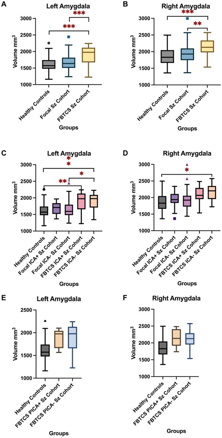
73 patients with only-focal seizures and 30 with FBTCS recorded during video EEG (VEEG) with respiratory monitoring were recruited prospectively during presurgical investigations. We acquired high-resolution T1-weighted anatomical and multi-shell diffusion images, and computed neurite orientation dispersion and density imaging (NODDI) metrics in all epilepsy patients and 69 healthy controls. Amygdala volumetric and microstructure alterations were compared between healthy subjects, and patients with only-focal seizures or FBTCS The FBTCS group was further subdivided by presence of ICA and PICA, verified by VEEG.
Bilateral amygdala volumes were significantly increased in the FBTCS cohort compared to healthy controls and the focal cohort. Patients with recorded PICA had the highest increase in bilateral amygdala volume of the FBTCS cohort.Amygdala neurite density index (NDI) values were significantly decreased in both the focal and FBTCS groups relative to healthy controls, with values in the FBTCS group being the lowest of the two. The presence of PICA was associated with significantly lower NDI values the non-apnea FBTCS group (p=0.004).
Individuals with FBTCS and PICA show significantly increased amygdala volumes and disrupted architecture bilaterally, with greater changes on the left side. The structural alterations reflected by NODDI and volume differences may be associated with inappropriate cardiorespiratory patterns mediated by the amygdala, particularly after FBTCS. Determination of amygdala volumetric and architectural changes may assist identification of individuals at risk.
癫痫性猝死(SUDEP)是癫痫患者的主要死因;然而,其病理生理学仍不清楚。局灶性至双侧强直阵挛发作(FBTCS)是一个主要危险因素,中枢介导的呼吸抑制可能会进一步增加风险。在此,我们确定了杏仁核的体积和微观结构,杏仁核是局灶性癫痫患者中可引发呼吸暂停的关键结构,并按是否存在FBTCS、发作期中枢性呼吸暂停(ICA)和发作后期中枢性呼吸暂停(PICA)进行分层。
在术前检查期间前瞻性招募了73例仅局灶性发作的患者和30例在视频脑电图(VEEG)监测呼吸时记录到FBTCS的患者。我们获取了高分辨率T1加权解剖图像和多壳扩散图像,并计算了所有癫痫患者和69名健康对照者的神经突方向离散度和密度成像(NODDI)指标。比较了健康受试者、仅局灶性发作患者或FBTCS患者之间杏仁核体积和微观结构的改变。FBTCS组根据VEEG证实的ICA和PICA的存在进一步细分。
与健康对照和局灶性发作组相比,FBTCS队列中的双侧杏仁核体积显著增加。记录到PICA的患者在FBTCS队列中的双侧杏仁核体积增加最大。相对于健康对照,局灶性发作组和FBTCS组的杏仁核神经突密度指数(NDI)值均显著降低,FBTCS组的值在两者中最低。PICA的存在与非呼吸暂停FBTCS组中显著更低的NDI值相关(p = 0.004)。
患有FBTCS和PICA的个体双侧杏仁核体积显著增加且结构破坏,左侧变化更大。NODDI反映的结构改变和体积差异可能与杏仁核介导的不适当心肺模式有关,尤其是在FBTCS之后。确定杏仁核体积和结构变化可能有助于识别有风险的个体。