Department of Radiology, Affiliated Nantong Hospital of Shanghai University (The Sixth People's Hospital of Nantong), Nantong, China.
Department of Radiology, Shanghai TCM-Integrated Hospital affiliated to Shanghai University of Traditional Chinese Medicine, Shanghai, China.
J Int Med Res. 2023 Mar;51(3):3000605231164005. doi: 10.1177/03000605231164005.
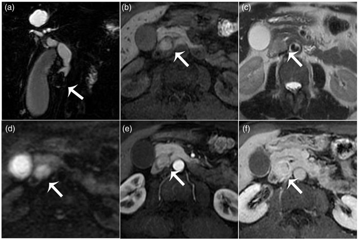
Primary biliary melanoma arises from proliferating melanocytes in the mucosal surface of the bile duct and is extremely rare. Since the vast majority of biliary melanomas represent metastases of cutaneous origin, accurate preoperative diagnosis of melanoma and exclusion of other primary sources are vital in cases involving primary lesions. Although melanomas with pigmented cells have typical signal characteristics, obtaining a non-invasive pre-treatment diagnosis remains difficult, due to their low incidence. Here, the case of a 61-year-old male Asian patient who presented with upper quadrant abdominal pain, swelling and jaundice for 2 weeks, and who was diagnosed with primary biliary melanoma following extensive preoperative blood analyses, computed tomography (CT) and magnetic resonance imaging (MRI), is described. Post-resection immunohistochemistry confirmed the diagnosis and the patient received six chemotherapy cycles of temozolomide and cisplatin, however, progression of multiple liver metastases was observed at the 18-month follow-up CT. The patient continued with pembrolizumab and died 17 months later. The present case of primary biliary melanoma is the first reported diagnosis based on typical MRI features and the exhaustive exclusion of a separate primary origin.
原发性胆管黑色素瘤由胆管黏膜表面增生的黑色素细胞引起,极为罕见。由于绝大多数胆管黑色素瘤是皮肤来源的转移瘤,因此对于涉及原发性病变的病例,准确的术前诊断黑色素瘤并排除其他原发性来源至关重要。尽管有色素细胞的黑色素瘤具有典型的信号特征,但由于其发病率低,因此获得非侵入性的术前诊断仍然具有挑战性。本文报道了一例 61 岁亚裔男性患者,其因上腹部疼痛、肿胀和黄疸持续 2 周就诊,经广泛的术前血液分析、计算机断层扫描(CT)和磁共振成像(MRI)检查后诊断为原发性胆管黑色素瘤。术后免疫组织化学检查证实了该诊断,患者接受了 6 个周期的替莫唑胺和顺铂化疗,但在 18 个月的 CT 随访中观察到多个肝转移灶进展。患者继续接受派姆单抗治疗,17 个月后死亡。本例原发性胆管黑色素瘤是首例基于典型 MRI 特征和详尽排除单独原发性起源的诊断。