Villanueva Pablo, Baldoncini Matías, Forlizzi Valeria, Campero Alvaro, Rangel Carlos Castillo, Granja Jaime Ordóñez, Sufianov Albert, Lucifero Alice Giotta, Luzzi Sabino
Department of Neurosurgery, Hospital Gobernador Ernesto Campos, Ushuaia, Tierra del Fuego, Argentina.
Laboratory of Microsurgical Neuroanatomy, Second Chair of Gross Anatomy, School of Medicine, University of Buenos Aires, Buenos Aires, Argentina.
Surg Neurol Int. 2023 Mar 24;14:97. doi: 10.25259/SNI_1095_2022. eCollection 2023.
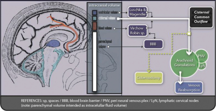
Cisternostomy is a surgical technique thought of and developed as an option for severe brain trauma treatment. It demands a particular knowledge and skill to microsurgically approach basal cisterns and effectively manipulate their contents. To perform this procedure safely, the anatomy and pathophysiology must be clearly understood.
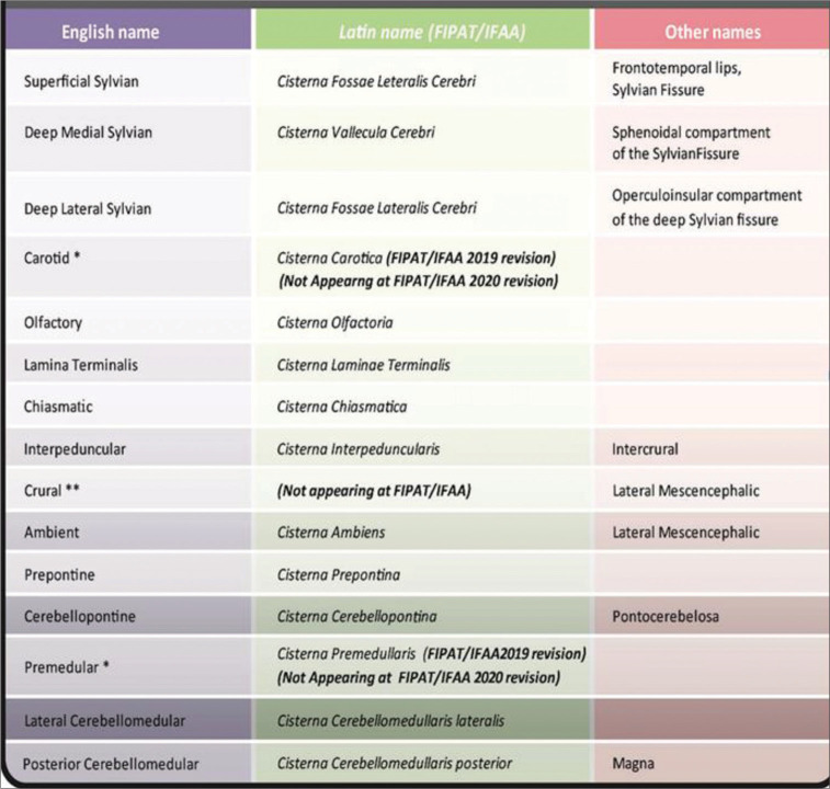
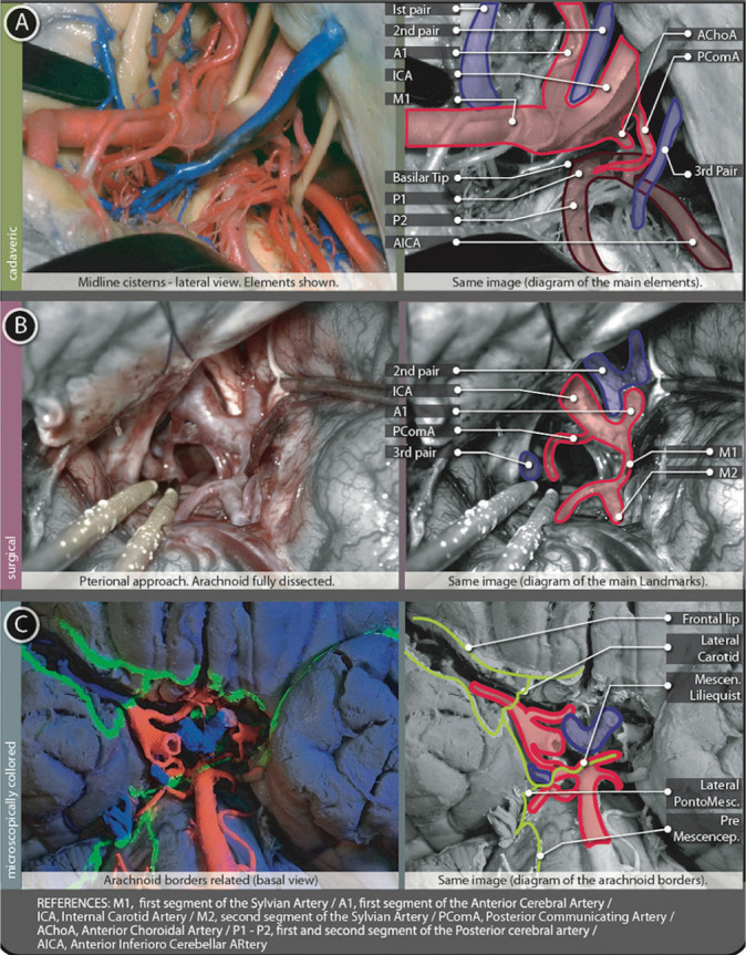
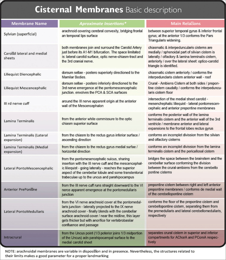
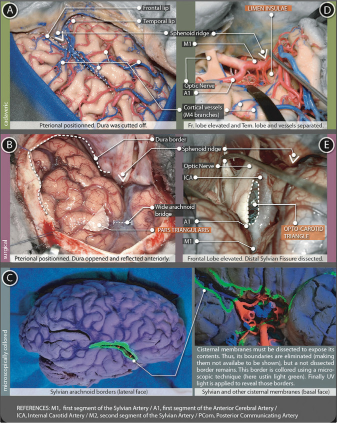
Detailed microscopic dissection and anatomical review were done, after a detailed reading of facts and recent publications about cisternostomy. Cisternal pathways and landmark planning are described and augmented using a new method to show de arachnoid borders. Finally, a brief discussion is written as a synopsis.
Cisternostomy requires thorough microscopic knowledge and microsurgical skills. This paper intends to provide information to understand better the anatomy related, thus, easing the learning curve. The technique used to show arachnoid borders, complementing cadaveric and surgical images, was useful for this purpose.
To perform this procedure safely, it is mandatory to handle microscopic details of cistern anatomy. Reaching a core cistern is necessary to assure effectiveness. This procedure needs, as well, surgical step-by-step landmark planning and performing. Cisternostomy could be a life-saving procedure and a new powerful tool for severe brain trauma treatment. Evidence is being collected to support its indications.
脑池造瘘术是一种作为严重脑外伤治疗选择而被构思和发展起来的外科技术。它需要特定的知识和技能来以显微外科方式接近脑基底池并有效操作其中的内容物。为了安全地实施该手术,必须清楚地了解其解剖结构和病理生理学。
在详细阅读有关脑池造瘘术的事实和近期出版物后,进行了详细的显微镜下解剖和解剖学回顾。使用一种显示蛛网膜边界的新方法描述并扩充了脑池路径和标志点规划。最后,撰写了一篇简短的讨论作为总结。
脑池造瘘术需要全面的显微镜知识和显微外科技能。本文旨在提供信息以更好地理解相关解剖结构,从而简化学习曲线。用于显示蛛网膜边界的技术,辅以尸体解剖和手术图像,为此目的很有用。
为了安全地实施该手术,必须掌握脑池解剖结构的微观细节。到达核心脑池对于确保有效性是必要的。该手术还需要逐步的手术标志点规划和实施。脑池造瘘术可能是一种挽救生命的手术,也是治疗严重脑外伤的一种新的有力工具。正在收集证据以支持其适应症。