Lebecq Alexis, Fangain Aurélie, Boussaroque Alice, Caillaud Marie-Cécile
Laboratoire Reproduction et Développement des Plantes, Université de Lyon, ENS de Lyon, UCB Lyon 1, CNRS, INRA, Lyon, France.
Quant Plant Biol. 2022 Feb 15;3:e4. doi: 10.1017/qpb.2022.1. eCollection 2022.
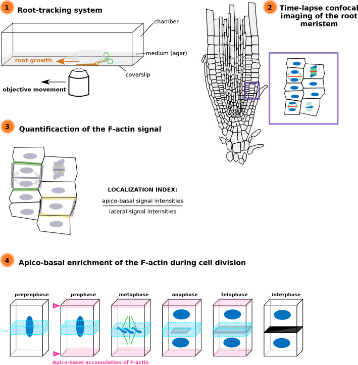
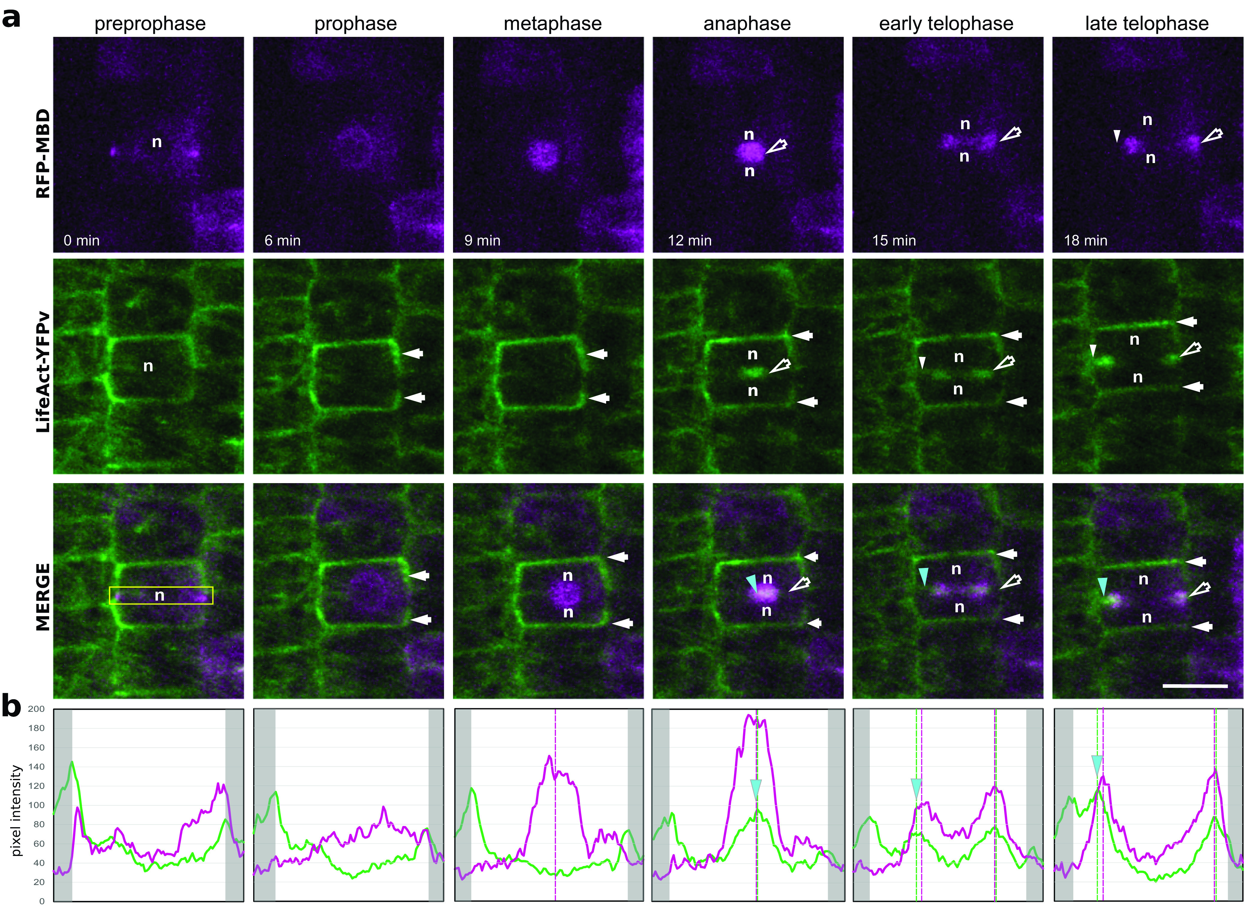
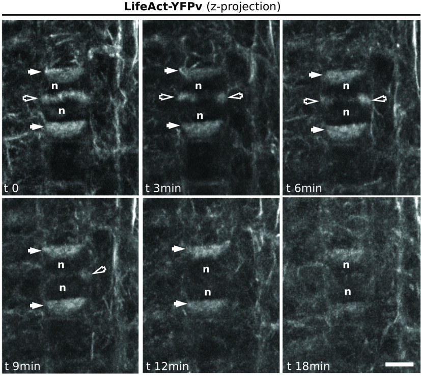
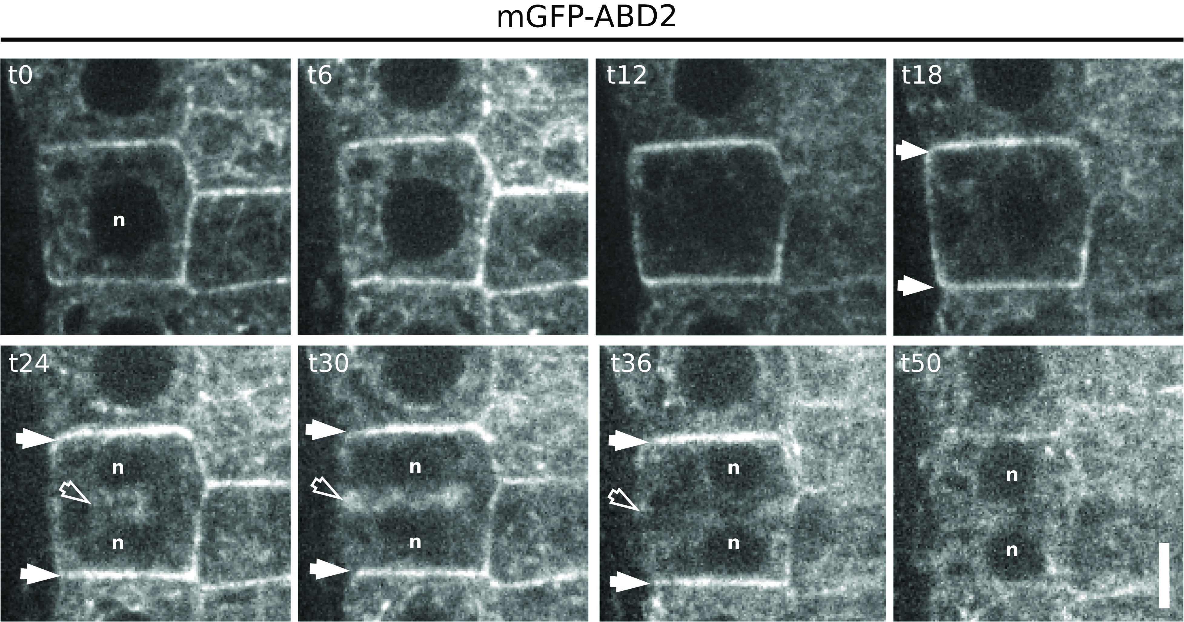
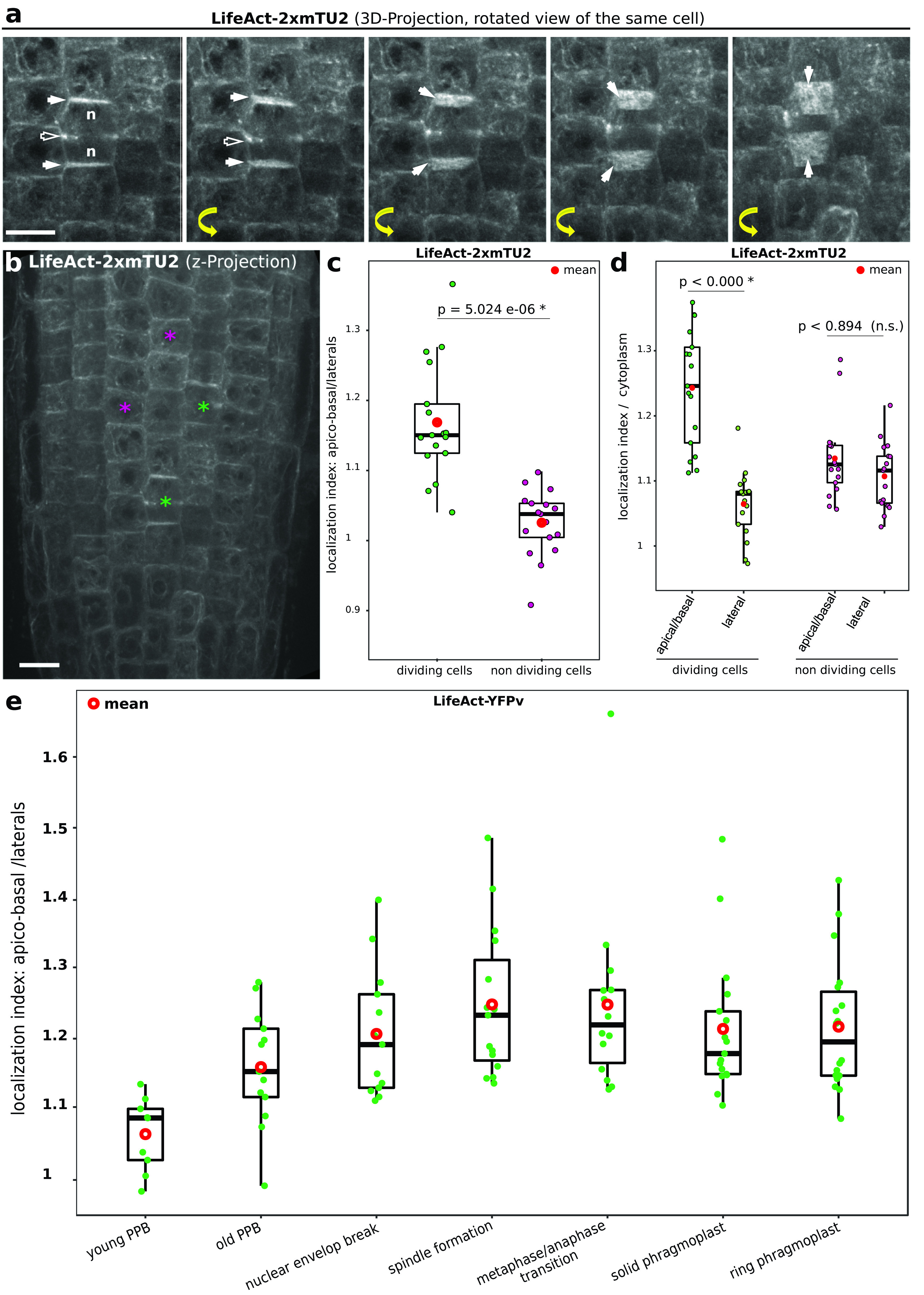
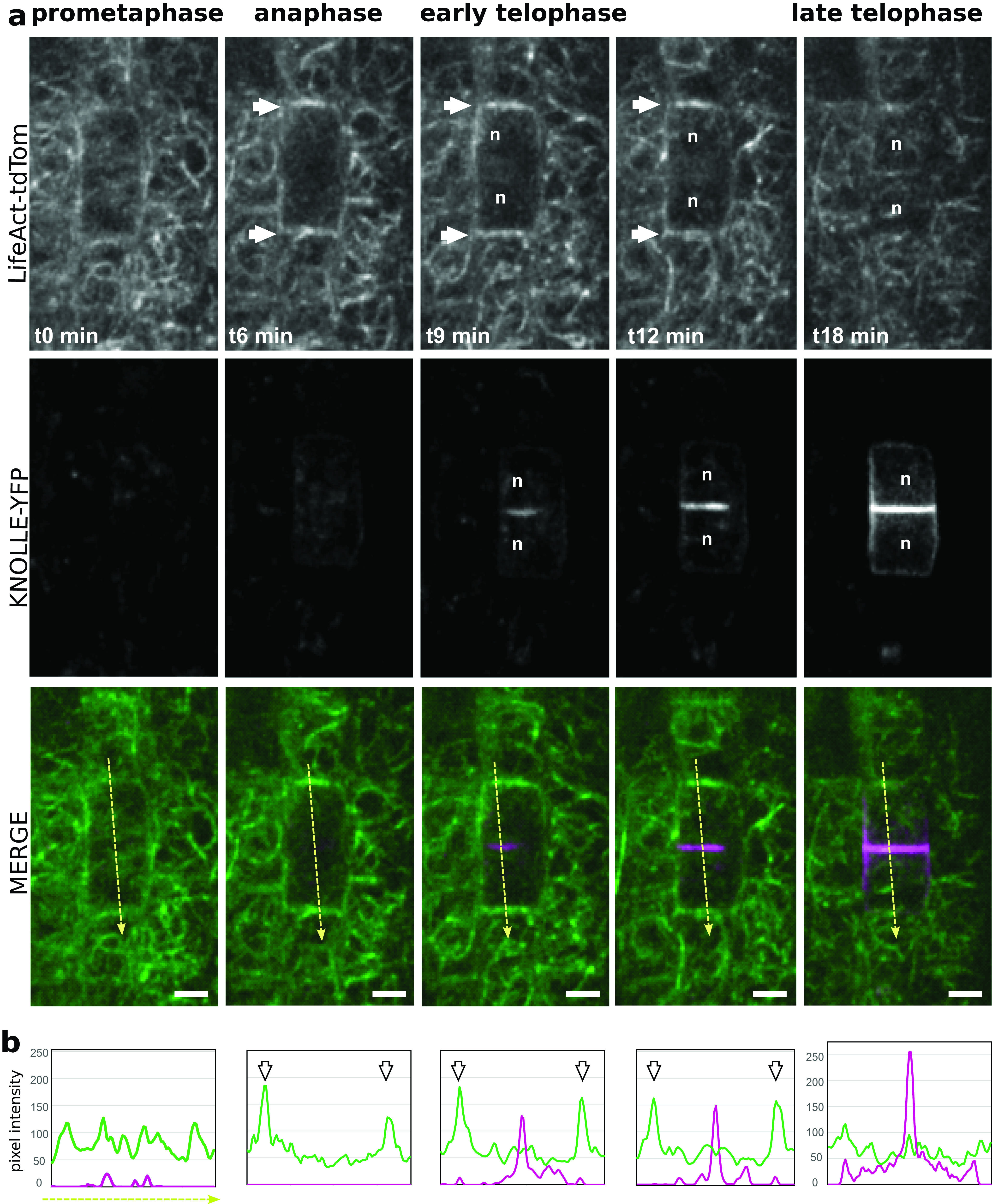
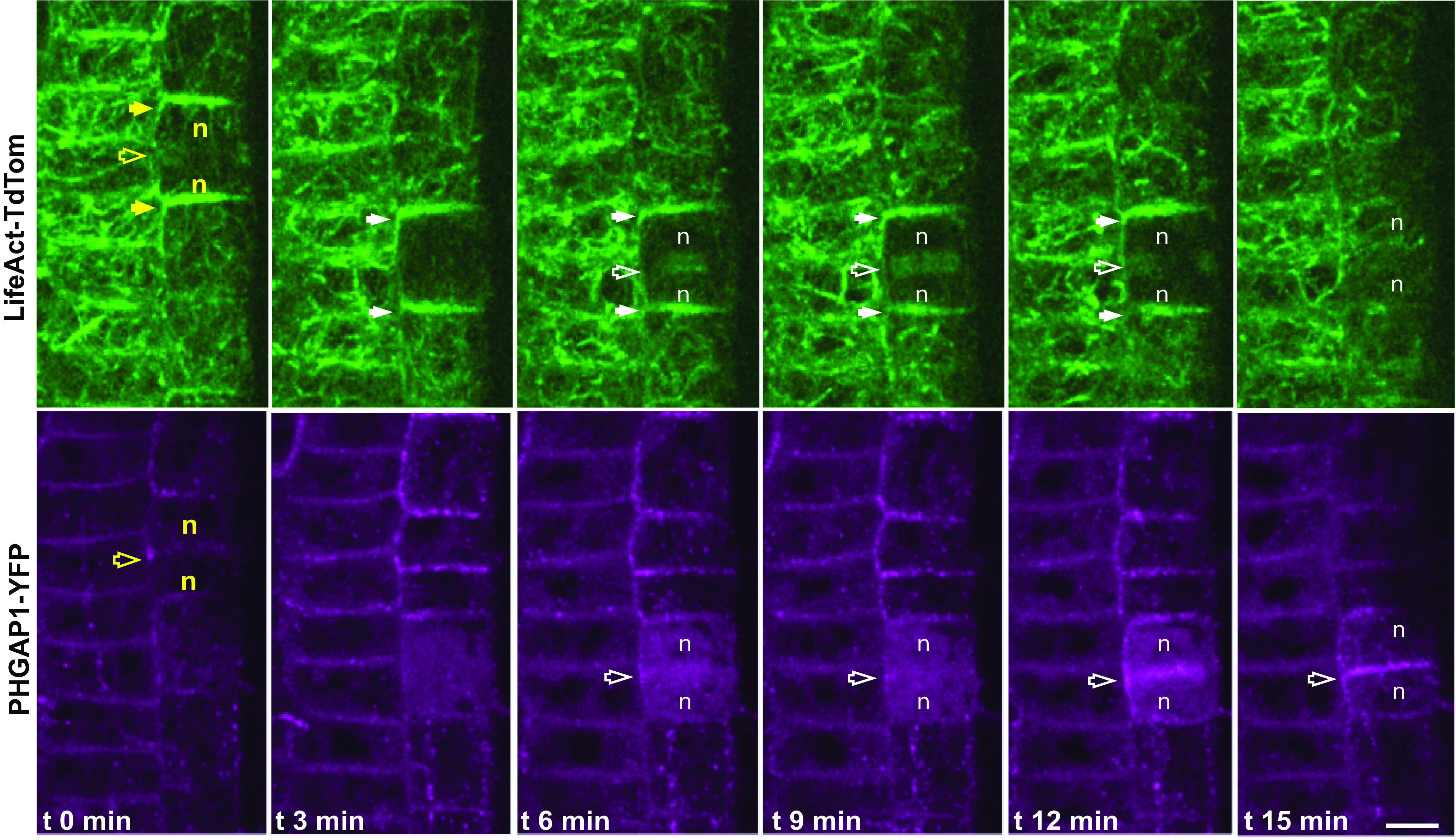
Cell division is a tightly regulated mechanism, notably in tissues where malfunctions can lead to tumour formation or developmental defects. This is particularly true in land plants, where cells cannot relocate and therefore cytokinesis determines tissue topology. In plants, cell division is executed in radically different manners than in animals, with the appearance of new structures and the disappearance of ancestral mechanisms. Whilst F-actin and microtubules closely co-exist, recent studies mainly focused on the involvement of microtubules in this key process. Here, we used a root tracking system to image the spatio-temporal dynamics of both F-actin reporters and cell division markers in dividing cells embedded in their tissues. In addition to the F-actin accumulation at the phragmoplast, we observed and quantified a dynamic apico-basal enrichment of F-actin from the prophase/metaphase transition until the end of the cytokinesis.
细胞分裂是一种受到严格调控的机制,在那些功能异常会导致肿瘤形成或发育缺陷的组织中尤为如此。在陆地植物中更是如此,因为植物细胞无法移动,所以胞质分裂决定了组织拓扑结构。在植物中,细胞分裂的执行方式与动物截然不同,会出现新的结构,同时一些原始机制会消失。虽然F-肌动蛋白和微管紧密共存,但最近的研究主要集中在微管在这一关键过程中的作用。在这里,我们使用了一个根系追踪系统,对嵌入组织中的分裂细胞中F-肌动蛋白报告基因和细胞分裂标记物的时空动态进行成像。除了在成膜体处积累F-肌动蛋白外,我们还观察并量化了从前期/中期过渡到胞质分裂结束时F-肌动蛋白在顶端-基部的动态富集。