Department of Systems Biology, Columbia University Irving Medical Center, New York, NY 10032, United States; Medical Scientist Training Program, Columbia University Irving Medical Center, New York, NY 10032, United States; Department of Psychiatry, Columbia University Irving Medical Center, New York, NY 10032, United States; Division of Molecular Therapeutics, New York State Psychiatric Institute, New York, NY 10032, United States.
Department of Psychiatry, Columbia University Irving Medical Center, New York, NY 10032, United States; Division of Molecular Therapeutics, New York State Psychiatric Institute, New York, NY 10032, United States.
Brain Behav Immun. 2023 Jul;111:277-291. doi: 10.1016/j.bbi.2023.04.008. Epub 2023 Apr 24.
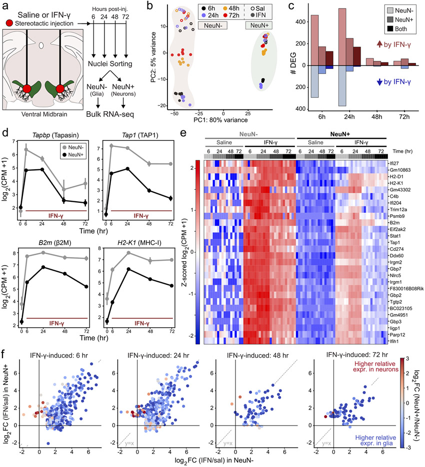
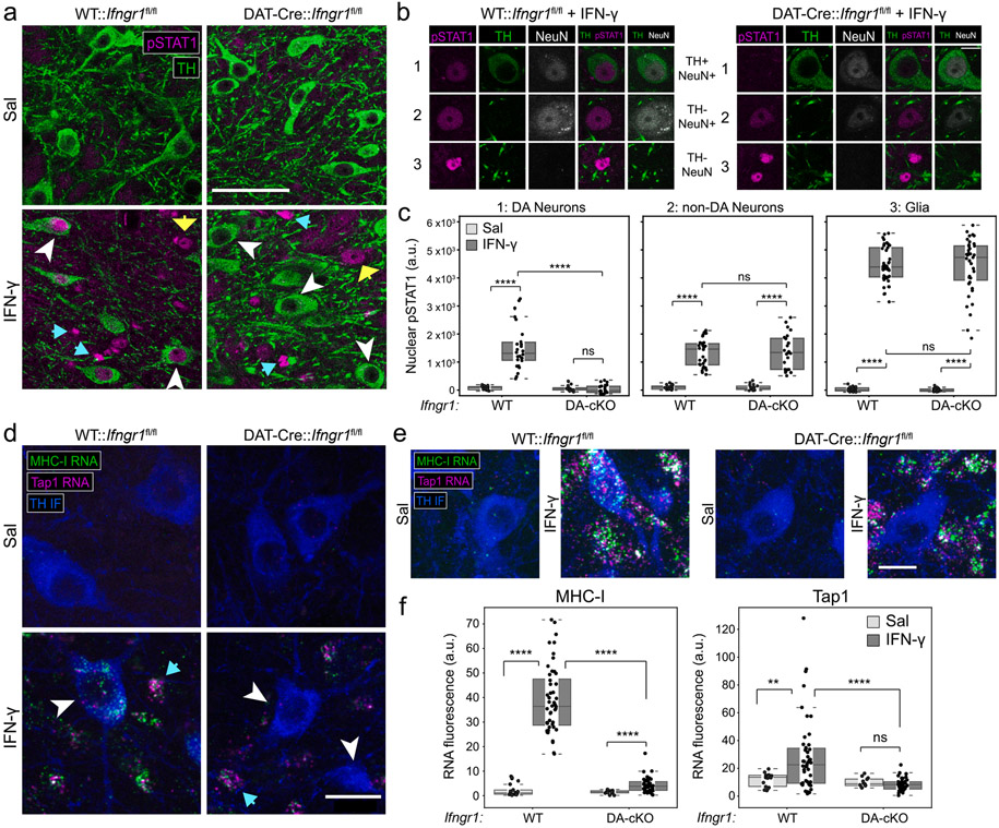
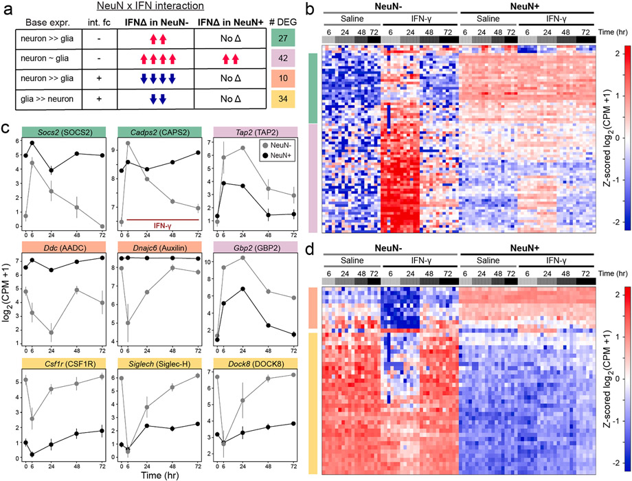
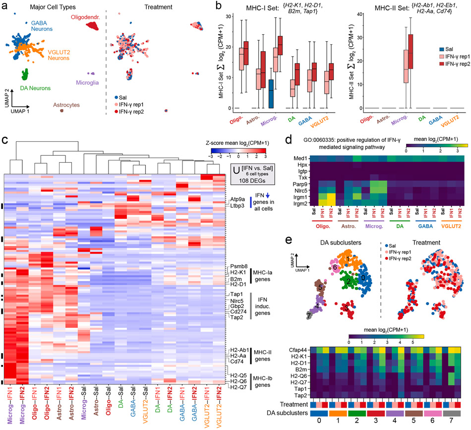
Dysregulated inflammation within the central nervous system (CNS) contributes to neuropathology in infectious, autoimmune, and neurodegenerative disease. With the exception of microglia, major histocompatibility complex (MHC) proteins are virtually undetectable in the mature, healthy central nervous system (CNS). Neurons have generally been considered incapable of antigen presentation, and although interferon gamma (IFN-γ) can elicit neuronal MHC class I (MHC-I) expression and antigen presentation in vitro, it has been unclear whether similar responses occur in vivo. Here we directly injected IFN-γ into the ventral midbrain of mature mice and analyzed gene expression profiles of specific CNS cell types. We found that IFN-γ upregulated MHC-I and associated mRNAs in ventral midbrain microglia, astrocytes, oligodendrocytes, and GABAergic, glutamatergic, and dopaminergic neurons. The core set of IFN-γ-induced genes and their response kinetics were similar in neurons and glia, but with a lower amplitude of expression in neurons. A diverse repertoire of genes was upregulated in glia, particularly microglia, which were the only cells to undergo cellular proliferation and express MHC classII (MHC-II) and associated genes. To determine if neurons respond directly via cell-autonomous IFN-γ receptor (IFNGR) signaling, we produced mutant mice with a deletion of the IFN-γ-binding domain of IFNGR1 in dopaminergic neurons, which resulted in a complete loss of dopaminergic neuronal responses to IFN-γ. Our results demonstrate that IFN-γ induces neuronal IFNGR signaling and upregulation of MHC-I and related genes in vivo, although the expression level is low compared to oligodendrocytes, astrocytes, and microglia.
中枢神经系统(CNS)内失调的炎症反应导致感染、自身免疫和神经退行性疾病的神经病理学。除了小胶质细胞之外,主要组织相容性复合体(MHC)蛋白在成熟、健康的中枢神经系统(CNS)中几乎检测不到。神经元通常被认为不能进行抗原呈递,尽管干扰素γ(IFN-γ)可以在体外诱导神经元 MHC 类 I(MHC-I)表达和抗原呈递,但尚不清楚是否在体内发生类似反应。在这里,我们直接将 IFN-γ注射到成熟小鼠的腹侧中脑,并分析特定 CNS 细胞类型的基因表达谱。我们发现,IFN-γ上调了腹侧中脑小胶质细胞、星形胶质细胞、少突胶质细胞以及 GABA 能、谷氨酸能和多巴胺能神经元中的 MHC-I 和相关 mRNA。IFN-γ诱导的基因及其反应动力学的核心集在神经元和神经胶质细胞中相似,但在神经元中的表达幅度较低。在神经胶质细胞中上调了多种基因,特别是小胶质细胞,小胶质细胞是唯一发生细胞增殖并表达 MHC 类 II(MHC-II)和相关基因的细胞。为了确定神经元是否通过细胞自主 IFN-γ受体(IFNGR)信号直接响应,我们在多巴胺能神经元中产生了 IFNGR1 的 IFN-γ结合域缺失的突变小鼠,这导致多巴胺能神经元对 IFN-γ的反应完全丧失。我们的结果表明,IFN-γ在体内诱导神经元 IFNGR 信号和 MHC-I 及相关基因的上调,尽管与少突胶质细胞、星形胶质细胞和小胶质细胞相比,其表达水平较低。