Department of Pediatrics, University of Chicago, Chicago, Illinois, USA.
Department of Physiology and Biophysics, Stony Brook University, Stony Brook, New York, USA.
J Biol Chem. 2023 Aug;299(8):104935. doi: 10.1016/j.jbc.2023.104935. Epub 2023 Jun 17.
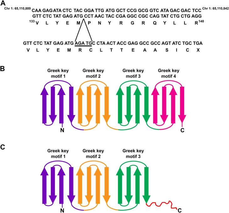
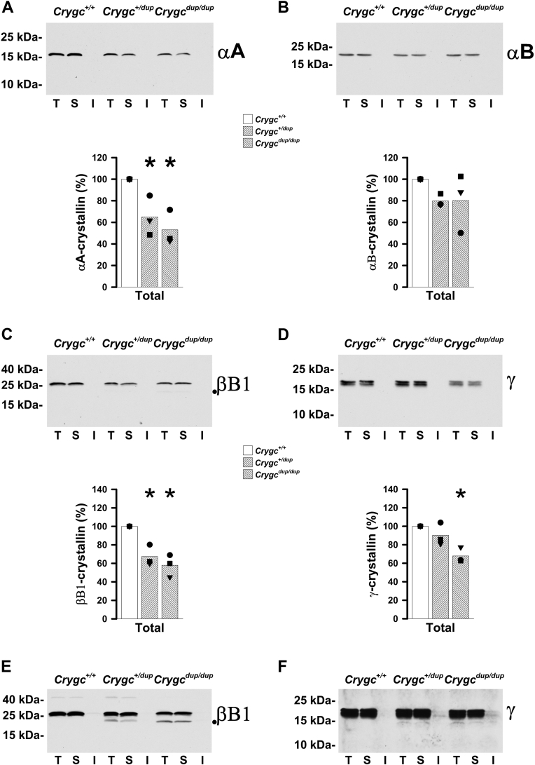
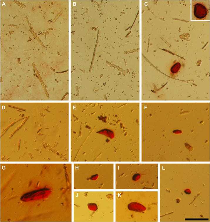
Connexin mutant mice develop cataracts containing calcium precipitates. To test whether pathologic mineralization is a general mechanism contributing to the disease, we characterized the lenses from a nonconnexin mutant mouse cataract model. By cosegregation of the phenotype with a satellite marker and genomic sequencing, we identified the mutant as a 5-bp duplication in the γC-crystallin gene (Crygc). Homozygous mice developed severe cataracts early, and heterozygous animals developed small cataracts later in life. Immunoblotting studies showed that the mutant lenses contained decreased levels of crystallins, connexin46, and connexin50 but increased levels of resident proteins of the nucleus, endoplasmic reticulum, and mitochondria. The reductions in fiber cell connexins were associated with a scarcity of gap junction punctae as detected by immunofluorescence and significant reductions in gap junction-mediated coupling between fiber cells in Crygc lenses. Particles that stained with the calcium deposit dye, Alizarin red, were abundant in the insoluble fraction from homozygous lenses but nearly absent in wild-type and heterozygous lens preparations. Whole-mount homozygous lenses were stained with Alizarin red in the cataract region. Mineralized material with a regional distribution similar to the cataract was detected in homozygous lenses (but not wild-type lenses) by micro-computed tomography. Attenuated total internal reflection Fourier-transform infrared microspectroscopy identified the mineral as apatite. These results are consistent with previous findings that loss of lens fiber cell gap junctional coupling leads to the formation of calcium precipitates. They also support the hypothesis that pathologic mineralization contributes to the formation of cataracts of different etiologies.
连接蛋白突变小鼠发生含有钙沉淀的白内障。为了测试病理性矿化是否是导致疾病的一般机制,我们对来自非连接蛋白突变小鼠白内障模型的晶状体进行了特征描述。通过表型与卫星标记的共分离和基因组测序,我们鉴定出突变体是γC-晶体蛋白(Crygc)基因中的 5 个碱基对重复。纯合子小鼠很早就发生严重白内障,杂合子动物在生命后期发展出小白内障。免疫印迹研究表明,突变体晶状体中晶体蛋白、连接蛋白 46 和连接蛋白 50 的水平降低,但核、内质网和线粒体的固有蛋白水平升高。纤维细胞连接蛋白的减少与免疫荧光检测到的间隙连接点状减少有关,并且 Crygc 晶状体中纤维细胞之间的间隙连接介导的偶联显著减少。用钙沉积染料茜素红染色的颗粒在纯合子晶状体的不溶性部分中丰富,但在野生型和杂合子晶状体制剂中几乎不存在。整个纯合子晶状体在白内障区域用茜素红染色。通过微计算机断层扫描在纯合子晶状体(但不在野生型晶状体)中检测到与白内障具有相似区域分布的矿化物质。衰减全内反射傅里叶变换红外微光谱鉴定出该矿物质为磷灰石。这些结果与先前的发现一致,即晶状体纤维细胞间隙连接偶联的丧失导致钙沉淀的形成。它们还支持病理性矿化有助于不同病因白内障形成的假说。