Department of Orthopedic Surgery, Nihon Koukan Hospital, 1-2-1 Koukandori, Kawasaki 210-0852, Japan.
Department of Orthopedic Surgery, Showa University School of Medicine, 1-5-8 Hatanodai, Shinagawa-ku, Tokyo 142-8666, Japan.
Medicina (Kaunas). 2023 Jun 1;59(6):1061. doi: 10.3390/medicina59061061.
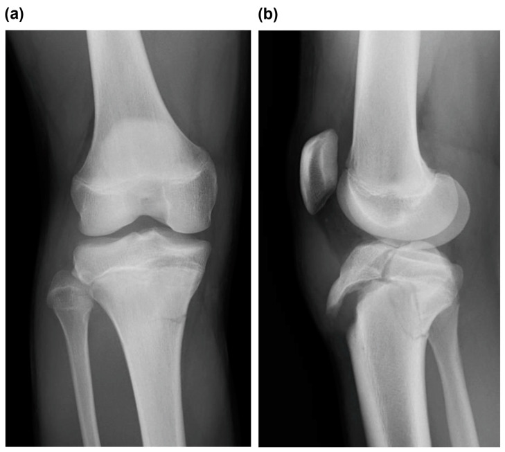
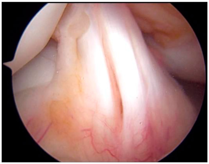
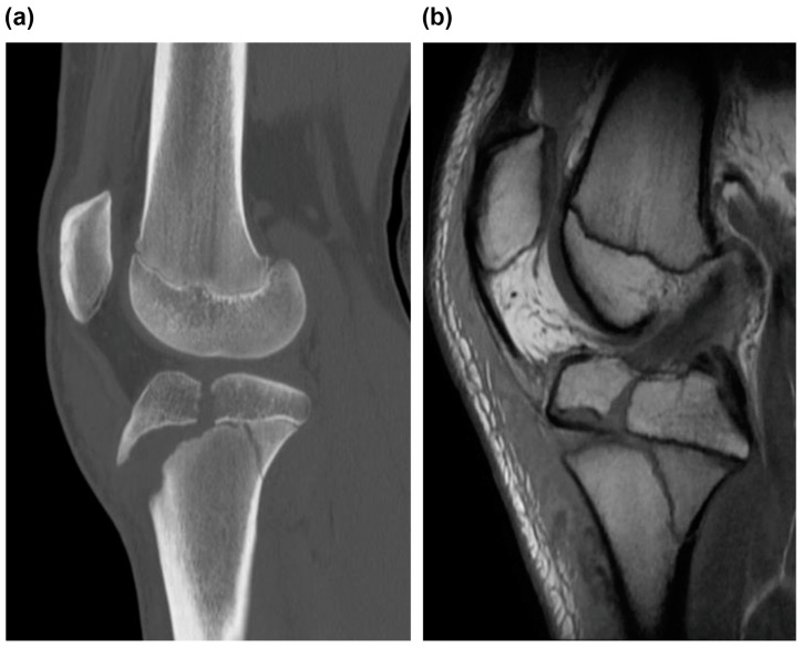
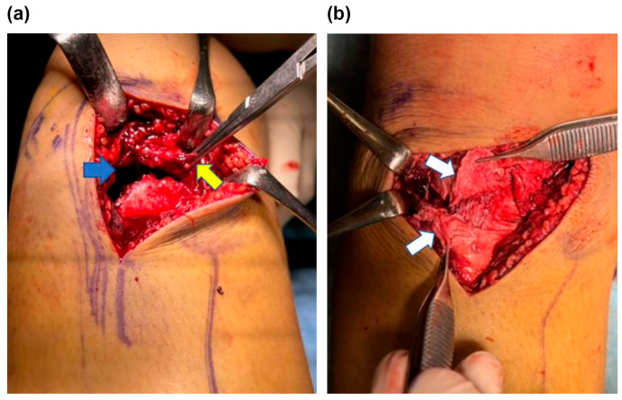
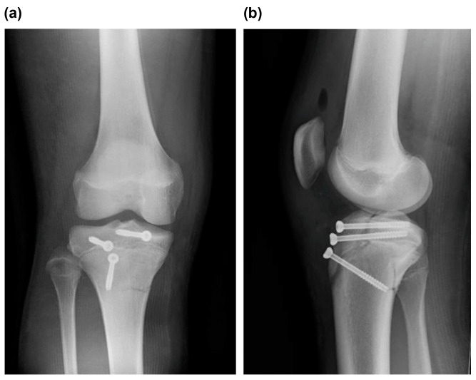
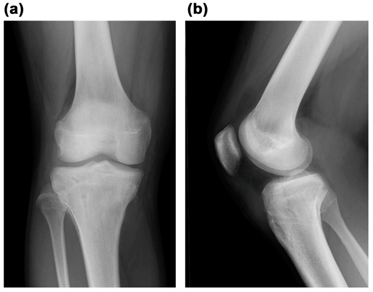
: Type V tibial tubercle avulsion fractures are extremely rare; therefore, information on them remains limited. Furthermore, although these fractures are intra-articular, to the best of our knowledge, there are no reports on their assessment via magnetic resonance imaging (MRI) or arthroscopy. Accordingly, this is the first report to describe the case of a patient undergoing detailed evaluation via MRI and arthroscopy. : A 13-year-old male adolescent athlete jumped while playing basketball, experienced discomfort and pain at the front of his knee, and fell down. He was transported to the emergency room by ambulance after he was unable to walk. The radiographic examination revealed a Type Ⅴ tibial tubercle avulsion fracture that was displaced. In addition, an MRI scan revealed a fracture line extending to the attachment of the anterior cruciate ligament (ACL); moreover, high MRI intensity and swelling due to ACL were observed, suggesting an ACL injury. On day 4 of the injury, open reduction and internal fixation were performed. Furthermore, 4 months after surgery, bone fusion was confirmed, and metal removal was performed. Simultaneously, an MRI scan obtained at the time of injury revealed findings suggestive of ACL injury; therefore, an arthroscopy was performed. Notably, no parenchymal ACL injury was observed, and the meniscus was intact. The patient returned to sports 6 months postoperatively. : Type V tibial tubercle avulsion fractures are known to be extremely rare. Based on our report, we suggest that MRI should be performed without hesitation if intra-articular injury is suspected.
V 型胫骨结节撕脱骨折极为罕见;因此,相关信息有限。此外,尽管这些骨折是关节内的,但据我们所知,尚无关于磁共振成像(MRI)或关节镜检查评估这些骨折的报告。因此,这是首次通过 MRI 和关节镜详细评估此类病例的报告。
一名 13 岁男性青少年运动员在打篮球时跳起,感到膝关节前部不适和疼痛,并摔倒在地。他无法行走后,被救护车送往急诊室。影像学检查显示 V 型胫骨结节撕脱骨折且有移位。此外,MRI 扫描显示骨折线延伸至前交叉韧带(ACL)的附着处;此外,由于 ACL 存在高 MRI 强度和肿胀,提示 ACL 损伤。伤后第 4 天行切开复位内固定术。术后 4 个月,确认骨融合,并进行金属取出术。同时,在受伤时进行的 MRI 扫描显示出 ACL 损伤的迹象;因此,进行了关节镜检查。值得注意的是,未观察到实质 ACL 损伤,半月板完整。术后 6 个月,患者恢复运动。
V 型胫骨结节撕脱骨折极为罕见。根据我们的报告,如果怀疑关节内损伤,我们建议毫不犹豫地进行 MRI 检查。