School of Medicine, Xiamen University, Xiamen, China.
Department of Radiology, The First Hospital of China Medical University, Shenyang, China.
CNS Neurosci Ther. 2024 Feb;30(2):e14363. doi: 10.1111/cns.14363. Epub 2023 Jul 19.
Acute kidney injury (AKI) has been associated with a variety of neurological problems, while the neurobiological mechanism remains unclear. In the present study, we utilized resting-state functional magnetic resonance imaging (rs-fMRI) to detect brain injury at an early stage and investigated the impact of microglia on the neuropathological mechanism of AKI.
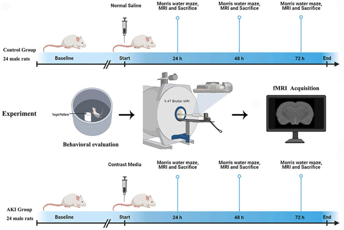
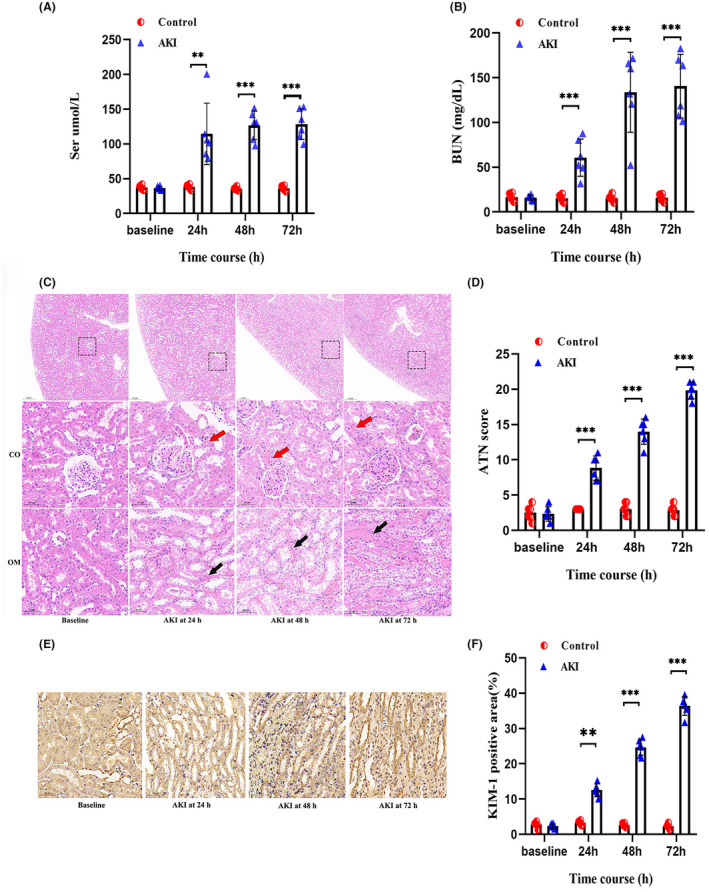
Rs-fMRI data were collected from AKI rats and the control group with a 9.4-Tesla scanner at 24, 48, and 72 h post administration of contrast medium or saline. The amplitude of low-frequency fluctuations (ALFF) was then compared across the groups at each time course. Additionally, flow cytometry and SMART-seq2 were employed to evaluate microglia. Furthermore, pathological staining and Western blot were used to analyze the samples.
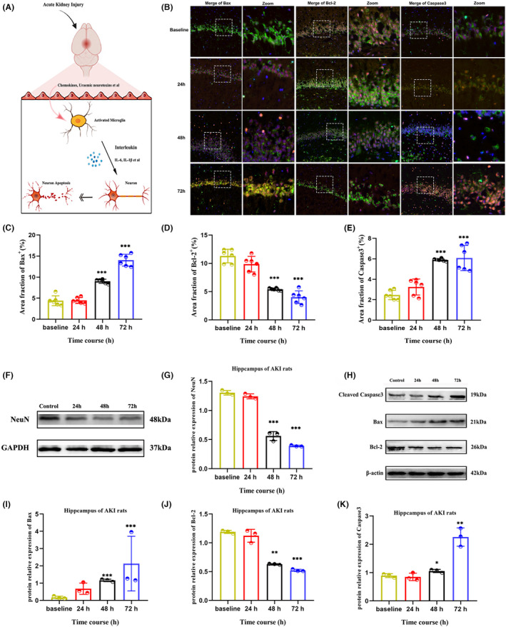
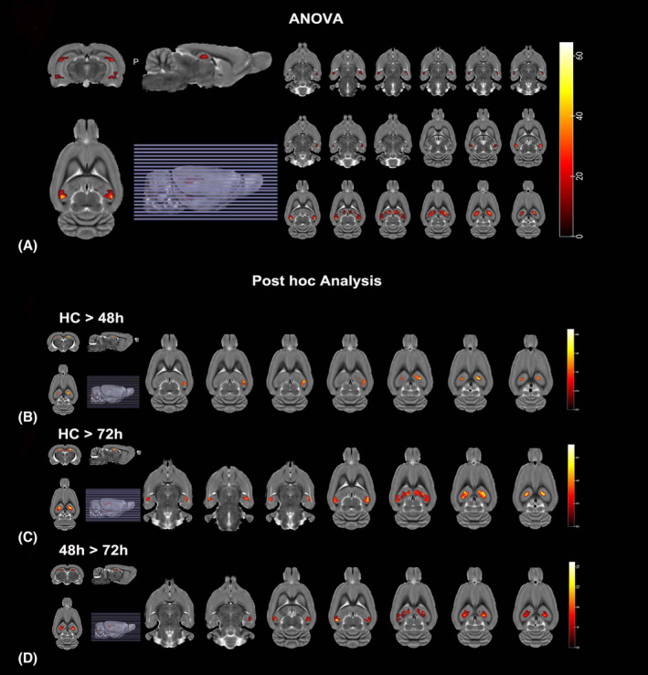
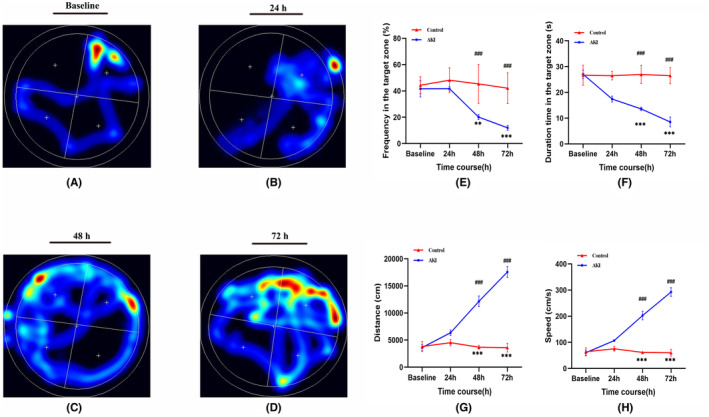
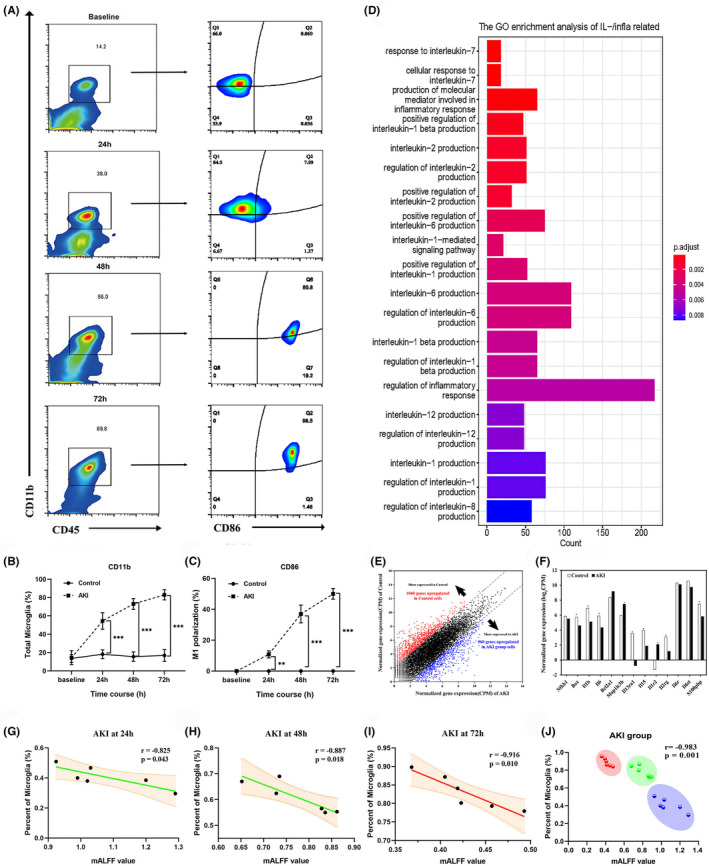
MRI results revealed that AKI led to a decreased ALFF in the hippocampus, particularly in the 48 h and 72 h groups. Additionally, western blot suggested that AKI-induced the neuronal apoptosis at 48 h and 72 h. Flow cytometry and confocal microscopy images demonstrated that AKI activated the aggregation of microglia into neurons at 24 h, with a strong upregulation of M1 polarization at 48 h and peaking at 72 h, accompanying with the release of proinflammatory cytokines. The ALFF value was strongly correlated with the proportion of microglia (|r| > 0.80, p < 0.001).
Our study demonstrated that microglia aggregation and inflammatory factor upregulation are significant mechanisms of AKI-induced neuronal apoptosis. We used fMRI to detect the alterations in hippocampal function, which may provide a noninvasive method for the early detection of brain injury after AKI.
急性肾损伤(AKI)与多种神经问题有关,但其神经生物学机制尚不清楚。本研究利用静息态功能磁共振成像(rs-fMRI)检测早期脑损伤,并探讨小胶质细胞对 AKI 神经病理机制的影响。
在 9.4T 扫描仪上对 AKI 大鼠和对照组进行 rs-fMRI 数据采集,在对比剂或生理盐水给药后 24、48 和 72 小时分别进行。然后比较各组在各时间点的低频振幅(ALFF)。此外,还采用流式细胞术和 SMART-seq2 评估小胶质细胞。进一步,采用病理染色和 Western blot 分析样本。
MRI 结果表明,AKI 导致海马区的 ALFF 降低,特别是在 48 小时和 72 小时组。此外,Western blot 表明 AKI 在 48 小时和 72 小时诱导神经元凋亡。流式细胞术和共聚焦显微镜图像显示,AKI 在 24 小时激活小胶质细胞向神经元聚集,48 小时 M1 极化强烈上调,72 小时达到高峰,同时释放促炎细胞因子。ALFF 值与小胶质细胞比例呈强相关(|r| > 0.80,p < 0.001)。
本研究表明,小胶质细胞聚集和炎症因子上调是 AKI 诱导神经元凋亡的重要机制。我们使用 fMRI 检测海马功能的改变,这可能为 AKI 后早期脑损伤提供一种非侵入性的检测方法。