• 文献检索
  • 文档翻译
  • 深度研究
  • 学术资讯
  • Suppr Zotero 插件Zotero 插件
  • 邀请有礼
  • 套餐&价格
  • 历史记录
应用&插件
Suppr Zotero 插件Zotero 插件浏览器插件Mac 客户端Windows 客户端微信小程序
定价
高级版会员购买积分包购买API积分包
服务
文献检索文档翻译深度研究API 文档MCP 服务
关于我们
关于 Suppr公司介绍联系我们用户协议隐私条款
关注我们

Suppr 超能文献

核心技术专利:CN118964589B侵权必究
粤ICP备2023148730 号-1Suppr @ 2026

文献检索

告别复杂PubMed语法,用中文像聊天一样搜索,搜遍4000万医学文献。AI智能推荐,让科研检索更轻松。

立即免费搜索

文件翻译

保留排版,准确专业,支持PDF/Word/PPT等文件格式,支持 12+语言互译。

免费翻译文档

深度研究

AI帮你快速写综述,25分钟生成高质量综述,智能提取关键信息,辅助科研写作。

立即免费体验

动脉自旋标记技术在评估横窦-乙状窦硬脑膜动静脉瘘治疗反应中的应用:一例报告

Arterial Spin Labeling Was Useful for Evaluating the Treatment Response of a Transverse-Sigmoid Sinus Dural Arteriovenous Fistula: A Case Report.

作者信息

Iwamura Masatoshi, Midorikawa Hiroshi, Kakuta Akihisa, Shibutani Koichi

机构信息

Department of Interventional Neuroradiology, Aomori Prefectural Central Hospital, Aomori, Aomori, Japan.

Department of Radiology, Aomori Prefectural Central Hospital, Aomori, Aomori, Japan.

出版信息

J Neuroendovasc Ther. 2021;15(2):113-119. doi: 10.5797/jnet.cr.2019-0119. Epub 2020 Sep 17.

DOI:10.5797/jnet.cr.2019-0119
PMID:37502803
原文链接:https://pmc.ncbi.nlm.nih.gov/articles/PMC10370806/
Abstract

OBJECTIVE

We report the case of a patient in whom arterial spin labeling (ASL) was useful for assessing the effects of treatment for a transverse-sigmoid sinus dural arteriovenous fistula (TSS-dAVF).

CASE PRESENTATION

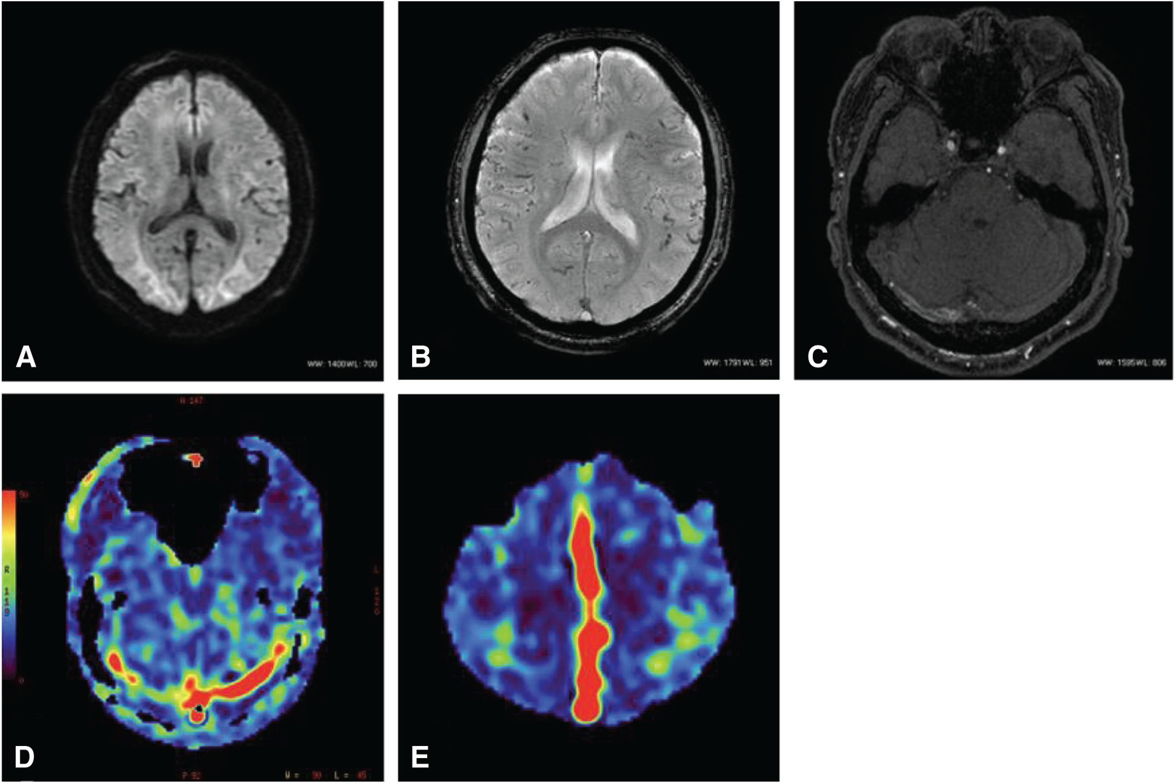
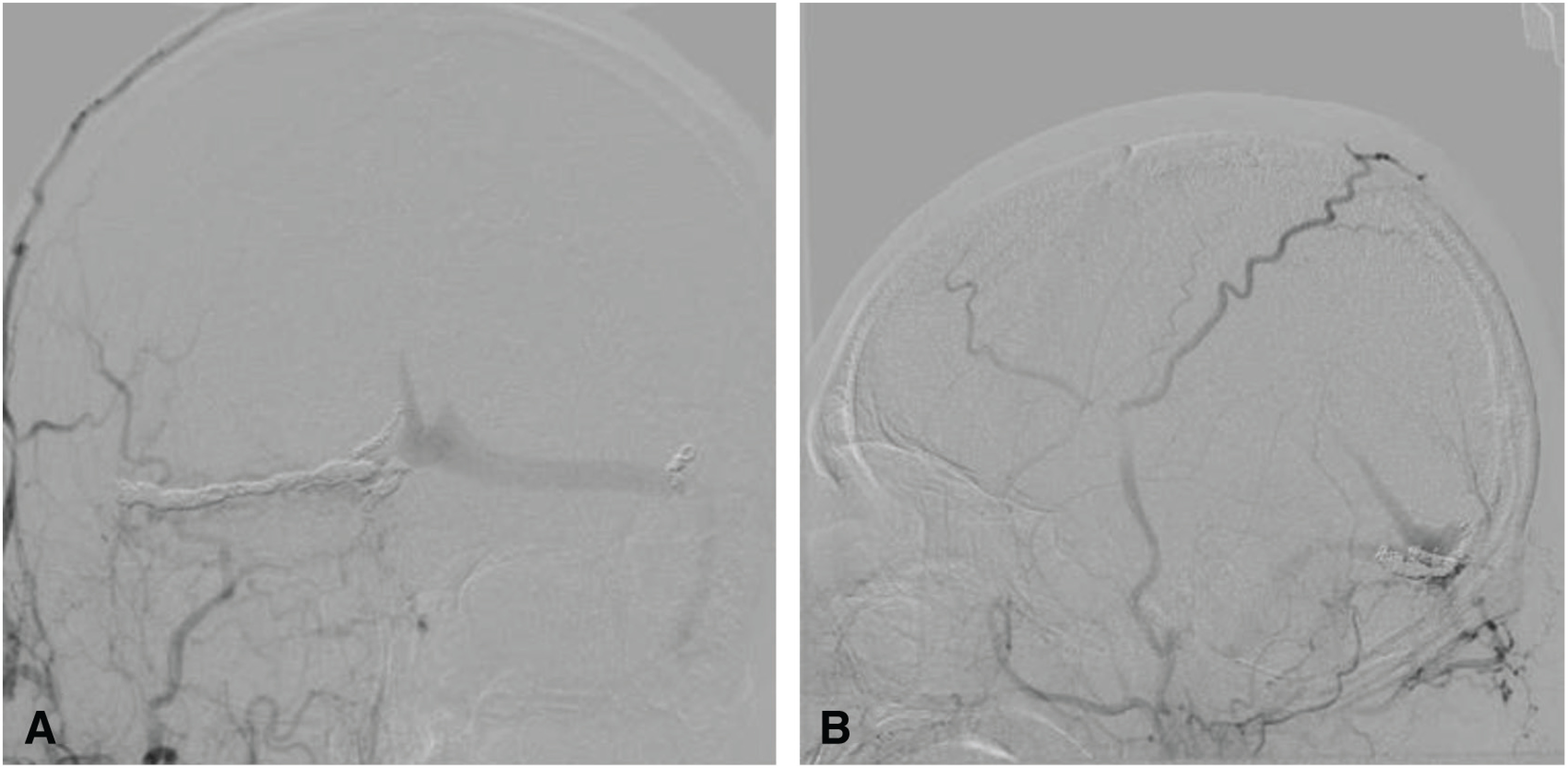
The patient was a 65-year-old man. Cerebral angiography demonstrated an aggressive dAVF involving the TSS, superior sagittal sinus (SSS), and the sinus confluence, with severe cortical and deep venous reflux. We performed multiple transarterial and transvenous embolizations for the TSS and sinus confluence lesion. The shunt disappeared almost completely after embolization. A high signal intensity that had been apparent in the SSS and straight sinus (StS) on ASL imaging before embolization disappeared after embolization. ASL imaging 3 months after embolization revealed slightly a high signal intensity in the StS, which was considered to be due to recurrence of the lesion. Moreover, recurrence of the confluence and TSS-dAVF was observed on cerebral angiography 6 months after embolization. As additional embolization was considered difficult, radiation therapy was recommended, but the patient refused; therefore, follow-up was performed. As ASL imaging findings were consistent with cerebral angiography findings, careful examination and monitoring of changes on ASL imaging were subsequently performed.

CONCLUSION

Follow-up using ASL imaging is useful to assess the effects of treatment performed for a dAVF.

摘要

目的

我们报告一例动脉自旋标记(ASL)有助于评估横窦-乙状窦硬脑膜动静脉瘘(TSS-dAVF)治疗效果的患者。

病例介绍

患者为一名65岁男性。脑血管造影显示为侵袭性硬脑膜动静脉瘘,累及横窦、上矢状窦(SSS)和窦汇,伴有严重的皮质和深部静脉逆流。我们对横窦和窦汇病变进行了多次经动脉和经静脉栓塞治疗。栓塞后分流几乎完全消失。栓塞前ASL成像中在SSS和直窦(StS)中明显的高信号强度在栓塞后消失。栓塞后3个月的ASL成像显示StS中有轻微的高信号强度,这被认为是由于病变复发所致。此外,栓塞后6个月的脑血管造影显示窦汇和TSS-dAVF复发。由于考虑再次栓塞困难,建议进行放射治疗,但患者拒绝;因此,进行了随访。由于ASL成像结果与脑血管造影结果一致,随后对ASL成像的变化进行了仔细检查和监测。

结论

使用ASL成像进行随访有助于评估对硬脑膜动静脉瘘所进行治疗的效果。

https://cdn.ncbi.nlm.nih.gov/pmc/blobs/7875/10370806/ded3bdb1375b/jnet-15-113-g006.jpg
https://cdn.ncbi.nlm.nih.gov/pmc/blobs/7875/10370806/3cf2de8c85ac/jnet-15-113-g001.jpg
https://cdn.ncbi.nlm.nih.gov/pmc/blobs/7875/10370806/8a6a3f0163bb/jnet-15-113-g002.jpg
https://cdn.ncbi.nlm.nih.gov/pmc/blobs/7875/10370806/9ec820f4c325/jnet-15-113-g003.jpg
https://cdn.ncbi.nlm.nih.gov/pmc/blobs/7875/10370806/26e073fe0fb1/jnet-15-113-g004.jpg
https://cdn.ncbi.nlm.nih.gov/pmc/blobs/7875/10370806/53cea4e10b15/jnet-15-113-g005.jpg
https://cdn.ncbi.nlm.nih.gov/pmc/blobs/7875/10370806/ded3bdb1375b/jnet-15-113-g006.jpg

相似文献

1
Arterial Spin Labeling Was Useful for Evaluating the Treatment Response of a Transverse-Sigmoid Sinus Dural Arteriovenous Fistula: A Case Report.动脉自旋标记技术在评估横窦-乙状窦硬脑膜动静脉瘘治疗反应中的应用:一例报告
J Neuroendovasc Ther. 2021;15(2):113-119. doi: 10.5797/jnet.cr.2019-0119. Epub 2020 Sep 17.
2
Fatal Hemorrhage Immediately after Transvenous Embolization of Transverse-sigmoid Sinus Dural Arteriovenous Fistulas: A Case Report.横窦-乙状窦窦硬膜动静脉瘘经静脉栓塞术后立即发生致命性出血:一例报告
J Neuroendovasc Ther. 2020;14(8):319-325. doi: 10.5797/jnet.cr.2019-0101. Epub 2020 Jun 5.
3
Single-session hematoma removal and transcortical venous approach for coil embolization of an isolated transverse-sigmoid sinus dural arteriovenous fistula in a hybrid operating room: illustrative case.在杂交手术室采用单期血肿清除及经皮质静脉入路对孤立性横窦-乙状窦硬膜动静脉瘘进行弹簧圈栓塞:病例报告
J Neurosurg Case Lessons. 2022 May 23;3(21):CASE2267. doi: 10.3171/CASE2267.
4
An Onyx tunnel: reconstructive transvenous balloon-assisted Onyx embolization for dural arteriovenous fistula of the transverse-sigmoid sinus.经静脉球囊辅助 Onyx 栓塞治疗横窦-乙状窦区硬脑膜动静脉瘘的 Onyx 隧道重建术。
J Neurosurg. 2018 Oct;129(4):922-927. doi: 10.3171/2017.5.JNS17287. Epub 2017 Nov 17.
5
A useful side-hole on a guiding catheter for transvenous embolization of a transverse-sigmoid sinus dural arteriovenous fistula.用于经静脉栓塞横窦-乙状窦硬膜动静脉瘘的引导导管上的一个有用的侧孔。
Neurosurgery. 2008 Jul;63(1 Suppl 1):ONSE91-2; discussion ONSE92. doi: 10.1227/01.neu.0000335019.06488.45.
6
Transverse-sigmoid Sinus Dural Arteriovenous Fistula Presenting with False Localizing Signs of Cavernous Sinus Dural Arteriovenous Fistula Due to Obstructed Cerebral Venous Outflow into the Bilateral Superior Ophthalmic Veins: A Case Report.横窦-乙状窦硬脑膜动静脉瘘因双侧眼上静脉脑静脉流出受阻出现海绵窦硬脑膜动静脉瘘假定位体征:一例报告
NMC Case Rep J. 2021 Nov 2;8(1):761-766. doi: 10.2176/nmccrj.cr.2021-0243. eCollection 2021.
7
[A Case of Cavernous Sinus Dural Arteriovenous Fistula after Transverse-Sigmoid Sinus Dural Arteriovenous Fistula Treatment with Radical Transvenous Coil Embolization].[经静脉根治性弹簧圈栓塞治疗横窦-乙状窦硬膜动静脉瘘后发生海绵窦硬膜动静脉瘘1例]
No Shinkei Geka. 2015 Aug;43(8):753-7. doi: 10.11477/mf.1436203114.
8
[Transvenous Embolization by Direct Puncture of the Superior Sagittal Sinus Using Indocyanine Green(ICG)Videoangiography for Treatment of Dural Arteriovenous Fistula of the Transverse-Sigmoid Sinus:A Case Report].[经静脉穿刺上矢状窦直接栓塞术联合吲哚菁绿(ICG)血管造影术治疗横窦-乙状窦硬脑膜动静脉瘘:1例报告]
No Shinkei Geka. 2017 Jul;45(7):591-598. doi: 10.11477/mf.1436203556.
9
Onyx removal after embolization of a superior sagittal sinus dural arteriovenous fistula involving scalp artery.涉及头皮动脉的上矢状窦硬脑膜动静脉瘘栓塞术后的Onyx移除。
Surg Neurol Int. 2016 Jun 3;7(Suppl 14):S410-4. doi: 10.4103/2152-7806.183518. eCollection 2016.
10
Occurrence of De Novo Dural Arteriovenous Fistula after Transvenous Embolization of Dural Arteriovenous Fistula : Case Reports of Two Patients.硬脑膜动静脉瘘经静脉栓塞术后新发硬脑膜动静脉瘘的发生:2例患者的病例报告
J Korean Neurosurg Soc. 2022 Jul;65(4):598-602. doi: 10.3340/jkns.2021.0144. Epub 2022 Apr 14.

引用本文的文献

1
Evaluating post-treatment residual intracranial arteriovenous shunting: a comparison of arterial spin labeling MRI and digital subtraction angiography.评估治疗后颅内残余动静脉分流:动脉自旋标记磁共振成像与数字减影血管造影的比较
Neuroradiology. 2025 May;67(5):1233-1250. doi: 10.1007/s00234-025-03548-7. Epub 2025 Feb 6.

本文引用的文献

1
Arterial spin labeling MR imaging aids to identify cortical venous drainage of dural arteriovenous fistulas.动脉自旋标记磁共振成像有助于识别硬脑膜动静脉瘘的皮质静脉引流。
Medicine (Baltimore). 2018 May;97(19):e0697. doi: 10.1097/MD.0000000000010697.
2
Arterial Spin-Labeling Improves Detection of Intracranial Dural Arteriovenous Fistulas with MRI.动脉自旋标记术提高 MRI 对颅内硬脑膜动静脉瘘的检出率。
AJNR Am J Neuroradiol. 2018 Apr;39(4):669-677. doi: 10.3174/ajnr.A5570. Epub 2018 Mar 15.
3
Epidemiologic Survey of Dural Arteriovenous Fistulas in Japan: Clinical Frequency and Present Status of Treatment.
日本硬脑膜动静脉瘘的流行病学调查:临床发病率及治疗现状
Acta Neurochir Suppl. 2016;123:185-8. doi: 10.1007/978-3-319-29887-0_26.
4
Detection of Cortical Venous Drainage and Determination of the Borden Type of Dural Arteriovenous Fistula by Means of 3D Pseudocontinuous Arterial Spin-Labeling MRI.通过三维伪连续动脉自旋标记磁共振成像检测硬脑膜动静脉瘘的皮质静脉引流并确定其博登分型
AJR Am J Roentgenol. 2016 Jul;207(1):163-9. doi: 10.2214/AJR.15.15171. Epub 2016 Apr 15.
5
Macrocyclic and Other Non-Group 1 Gadolinium Contrast Agents Deposit Low Levels of Gadolinium in Brain and Bone Tissue: Preliminary Results From 9 Patients With Normal Renal Function.大环类及其他非1类钆对比剂在脑和骨组织中钆沉积水平低:9例肾功能正常患者的初步结果
Invest Radiol. 2016 Jul;51(7):447-53. doi: 10.1097/RLI.0000000000000252.
6
Epidemiology of dural arteriovenous fistula in Japan: Analysis of Japanese Registry of Neuroendovascular Therapy (JR-NET2).日本硬脑膜动静脉瘘的流行病学:日本神经血管内治疗注册研究(JR-NET2)分析
Neurol Med Chir (Tokyo). 2014;54(1):63-71. doi: 10.2176/nmc.st.2013-0172. Epub 2013 Dec 27.
7
Perfusion imaging of brain tumors using arterial spin-labeling: correlation with histopathologic vascular density.利用动脉自旋标记技术对脑肿瘤进行灌注成像:与组织病理学血管密度的相关性
AJNR Am J Neuroradiol. 2008 Apr;29(4):688-93. doi: 10.3174/ajnr.A0903. Epub 2008 Jan 9.
8
Pulsed arterial spin labeling parameter optimization for an elderly population.老年人群的脉冲动脉自旋标记参数优化
J Magn Reson Imaging. 2006 Mar;23(3):398-403. doi: 10.1002/jmri.20503.
9
Intracranial dural arteriovenous fistulas: evaluation with combined 3D time-of-flight MR angiography and MR digital subtraction angiography.颅内硬脑膜动静脉瘘:联合三维时间飞跃磁共振血管造影和磁共振数字减影血管造影评估
AJR Am J Roentgenol. 2004 Jan;182(1):183-90. doi: 10.2214/ajr.182.1.1820183.
10
Pediatric perfusion imaging using pulsed arterial spin labeling.使用脉冲动脉自旋标记的儿科灌注成像。
J Magn Reson Imaging. 2003 Oct;18(4):404-13. doi: 10.1002/jmri.10372.