The State Key Laboratory of Oral & Maxillofacial Reconstruction and Regeneration, Key Laboratory of Oral Biomedicine Ministry of Education, Hubei Key Laboratory of Stomatology, School & Hospital of Stomatology, Wuhan University, Wuhan, China.
Department of Cariology and Endodontics, School and Hospital of Stomatology, Wuhan University, Wuhan, China.
Clin Oral Investig. 2023 Sep;27(9):5317-5329. doi: 10.1007/s00784-023-05152-6. Epub 2023 Aug 2.
This study was aimed at evaluating the clinical and radiological outcomes of novel dynamic navigation (DN)-aided endodontic microsurgery (EMS), with an analysis of potential prognostic factors.
Forty-six teeth from 32 patients who received DN-aided EMS were included. Clinical and radiographic assessments were performed at least 1 year postoperatively. Two calibrated endodontists assessed radiological outcomes according to two-dimensional (2D) periapical radiography (PA) and three-dimensional (3D) cone-beam computed tomography (CBCT) imaging using Rud's and Molven's criteria and modified PENN 3D criteria, respectively. Fisher's exact test was used for statistical analysis of the predisposing factors.
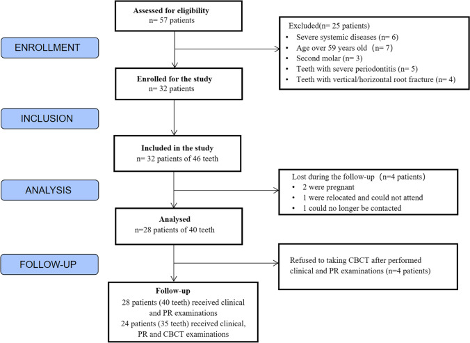
Of the 32 patients with 46 treated teeth, 28 with 40 teeth were available for follow-up. Of the 28 patients, four (five teeth) refused to undergo CBCT and only underwent clinical and PA examinations, and the remaining 24 (35 teeth) underwent clinical, PA, and CBCT examinations. Combined clinical and radiographic data revealed a 95% (38/40) success rate in 2D healing evaluations and a 94.3% (33/35) success rate in 3D healing evaluations. No significant effect was found in sex, age, tooth type, arch type, preoperative lesion volume, preoperative maximum lesion size, presence/absence of crown and post, and the root canal filling state on the outcome of DN-aided EMS.
DN-aided EMS has a favorable prognosis and could be considered an effective and reliable treatment strategy. Further investigations with larger sample sizes are required to confirm these results.
DN-aided EMS could be considered an effective and reliable treatment strategy.
本研究旨在评估新型动态导航(DN)辅助根管内显微手术(EMS)的临床和影像学效果,并分析潜在的预后因素。
纳入 32 例患者的 46 颗接受 DN 辅助 EMS 治疗的患牙。术后至少 1 年进行临床和影像学评估。2 位经过校准的牙髓病专家分别使用 Rud 和 Molven 标准以及改良的 PENN 3D 标准,基于二维(2D)根尖片(PA)和三维(3D)锥形束 CT(CBCT)影像,对放射学结果进行评估。采用 Fisher 确切检验对潜在预后因素进行统计学分析。
32 例患者(46 颗患牙)中,28 例(40 颗患牙)患者获得随访。28 例患者中,有 4 例(5 颗牙)拒绝接受 CBCT 检查,仅接受临床和 PA 检查,其余 24 例(35 颗牙)接受了临床、PA 和 CBCT 检查。联合临床和放射学数据显示,2D 愈合评估的成功率为 95%(38/40),3D 愈合评估的成功率为 94.3%(33/35)。性别、年龄、牙位、牙弓类型、术前病变体积、术前最大病变尺寸、有无冠和桩以及根管充填状态对 DN 辅助 EMS 的效果无显著影响。
DN 辅助 EMS 预后良好,可被视为一种有效且可靠的治疗策略。需要进一步开展更大样本量的研究以证实这些结果。
DN 辅助 EMS 可被视为一种有效且可靠的治疗策略。