Ghafar Norzana Abd, Jalil Nahdia Afiifah Abdul, Kamarudin Taty Anna
Pusat Perubatan Universiti Kebangsaan Malaysia, 56000 Cheras, Kuala Lumpur, Malaysia.
Department of Anatomy, Faculty of Medicine, Universiti Kebangsaan Malaysia, 56000 Cheras, Kuala Lumpur, Malaysia.
Asian Biomed (Res Rev News). 2021 Oct 29;15(5):199-212. doi: 10.2478/abm-2021-0026. eCollection 2021 Oct.
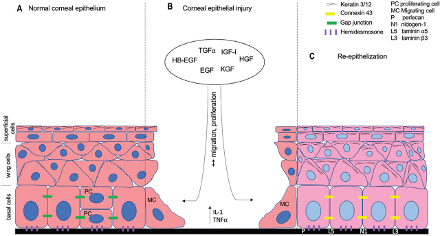
The corneal epithelium (CE) forms the outermost layer of the cornea. Despite its thickness of only 50 μm, the CE plays a key role as an initial barrier against any insults to the eye and contributes to the light refraction onto the retina required for clear vision. In the event of an injury, the cornea is equipped with many strategies contributing to competent wound healing, including angiogenic and immune privileges, and mechanotransduction. Various factors, including growth factors, keratin, cytokines, integrins, crystallins, basement membrane, and gap junction proteins are involved in CE wound healing and serve as markers in the healing process. Studies of CE wound healing are advancing rapidly in tandem with the rise of corneal bioengineering, which employs limbal epithelial stem cells as the primary source of cells utilizing various types of biomaterials as substrates.
角膜上皮(CE)构成角膜的最外层。尽管其厚度仅为50微米,但CE作为抵御眼部任何损伤的初始屏障发挥着关键作用,并有助于将光线折射到视网膜上以实现清晰视力。在发生损伤时,角膜具备多种有助于有效伤口愈合的机制,包括血管生成和免疫特权以及机械转导。多种因素,包括生长因子、角蛋白、细胞因子、整合素、晶状体蛋白、基底膜和缝隙连接蛋白,都参与了角膜上皮伤口愈合,并在愈合过程中作为标志物。随着角膜生物工程的兴起,角膜上皮伤口愈合的研究也在迅速发展,角膜生物工程利用角膜缘上皮干细胞作为主要细胞来源,并使用各种类型的生物材料作为基质。