Du Miao, Chen Shikun, Chen Yang, Yuan Xinxu, Dong Huansheng
College of Animal Science and Technology, Qingdao Agricultural University, Qingdao 266109, China.
College of Veterinary Medicine, Murdoch University, Murdoch, Western Australia 6150, Australia.
Anim Biosci. 2024 Jan;37(1):50-60. doi: 10.5713/ab.23.0175. Epub 2023 Aug 28.
Testicular fat deposition has been reported to affect animal reproduction. However, the underlying mechanism remains poorly understood. The present study explored whether sperm meiosis and testosterone synthesis contribute to mouse testicular fat depositioninduced reproductive performance.
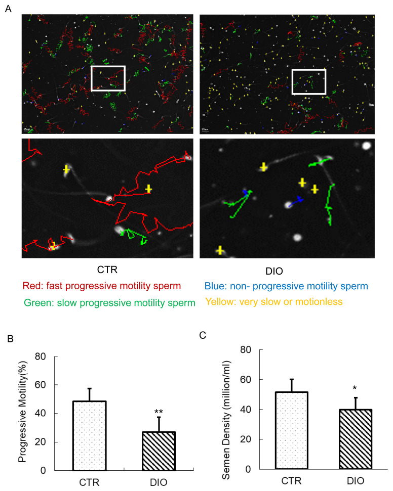
High fat diet (HFD)-induced obesity CD1 mice (DIO) were used as a testicular fat deposition model. The serum hormone test was performed by agent kit. The quality of sperm was assessed using a Sperm Class Analyzer. Testicular tissue morphology was analyzed by histochemical methods. The expression of spermatocyte marker molecules was monitored by an immuno-fluorescence microscope during meiosis. Analysis of the synthesis of testosterone was performed by real-time polymerase chain reaction and reagent kit.
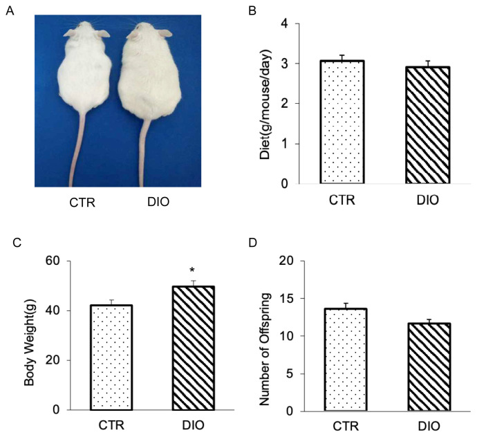
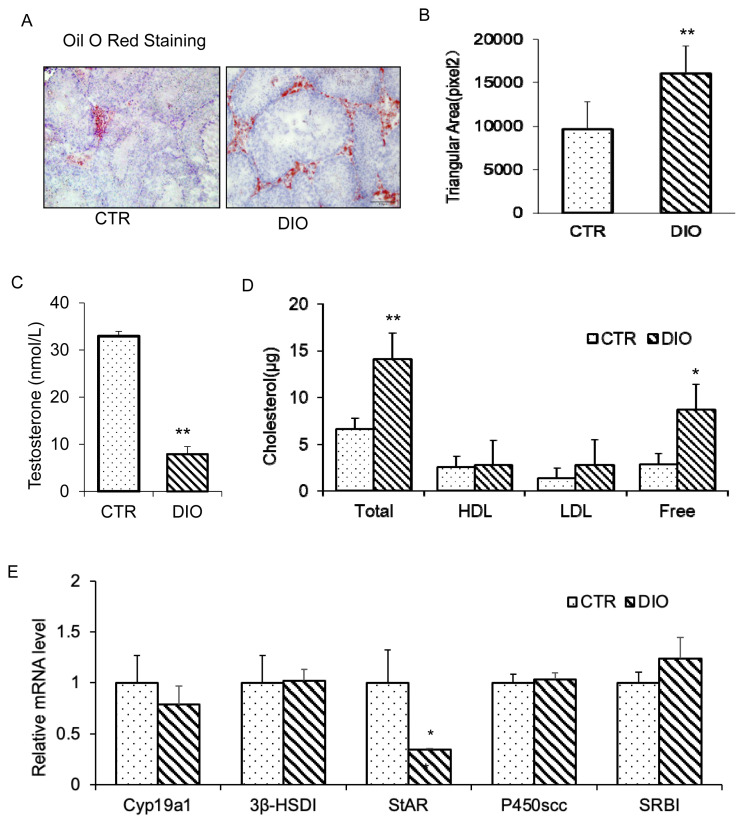
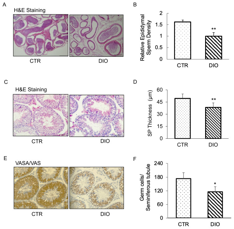
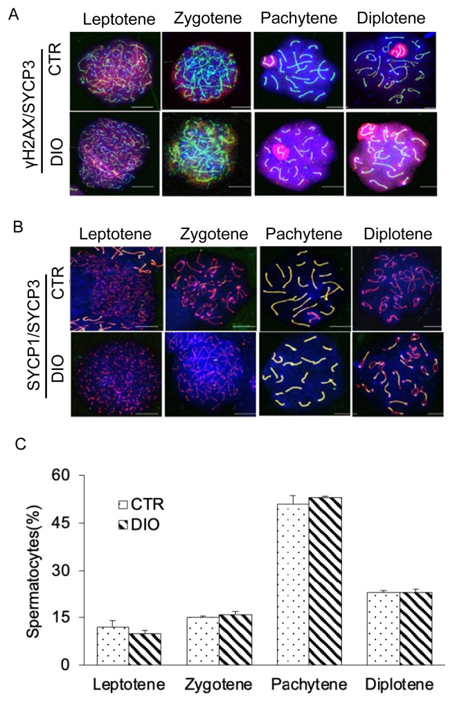
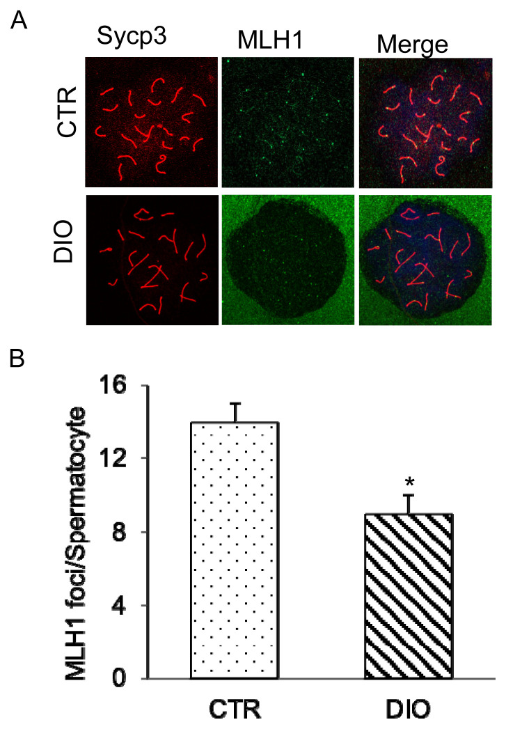
It was found that there was a significant increase in body weight among DIO mice, however, the food intake showed no difference compared to control mice fed a normal diet (CTR). The number of offspring in DIO mice decreased, but there was no significant difference from the CTR group. The levels of follicle-stimulating hormone were lower in DIO mice and their luteinizing hormone levels were similar. The results showed a remarkable decrease in sperm density and motility among DIO mice. We also found that fat accumulation affected the meiosis process, mainly reflected in the cross-exchange of homologous chromosomes. In addition, overweight increased fat deposition in the testis and reduced the expression of testosterone synthesis-related enzymes, thereby affecting the synthesis and secretion of testosterone by testicular Leydig cells.
Fat accumulation in the testes causes testicular cell dysfunction, which affects testosterone hormone synthesis and ultimately affects sperm formation.
据报道,睾丸脂肪沉积会影响动物繁殖。然而,其潜在机制仍知之甚少。本研究探讨精子减数分裂和睾酮合成是否对小鼠睾丸脂肪沉积诱导的生殖性能有影响。
采用高脂饮食(HFD)诱导的肥胖CD1小鼠(DIO)作为睾丸脂肪沉积模型。使用试剂盒进行血清激素检测。使用精子分类分析仪评估精子质量。通过组织化学方法分析睾丸组织形态。在减数分裂期间,通过免疫荧光显微镜监测精母细胞标记分子的表达。通过实时聚合酶链反应和试剂盒进行睾酮合成分析。
发现DIO小鼠体重显著增加,然而,与喂食正常饮食的对照小鼠(CTR)相比,食物摄入量没有差异。DIO小鼠的后代数量减少,但与CTR组没有显著差异。DIO小鼠的促卵泡激素水平较低,其黄体生成素水平相似。结果显示DIO小鼠的精子密度和活力显著降低。我们还发现脂肪堆积影响减数分裂过程,主要体现在同源染色体的交叉互换上。此外,超重增加了睾丸中的脂肪沉积,并降低了睾酮合成相关酶的表达,从而影响睾丸间质细胞睾酮的合成和分泌。
睾丸中的脂肪堆积会导致睾丸细胞功能障碍,影响睾酮激素合成,最终影响精子形成。