Department of Pathology and Laboratory Medicine, School of Medicine, Indiana University, Indianapolis, IN, 46202, USA.
Division of Neurology, University of Campania "Luigi Vanvitelli", Caserta, Italy.
Acta Neuropathol Commun. 2023 Aug 31;11(1):141. doi: 10.1186/s40478-023-01631-9.
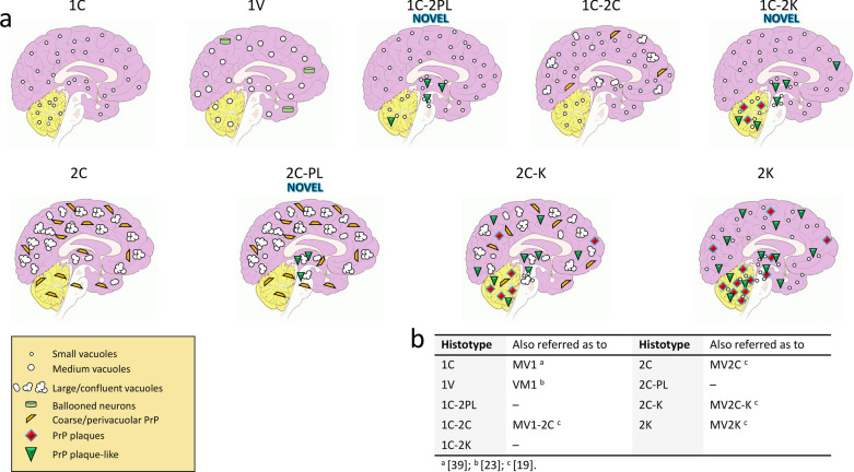
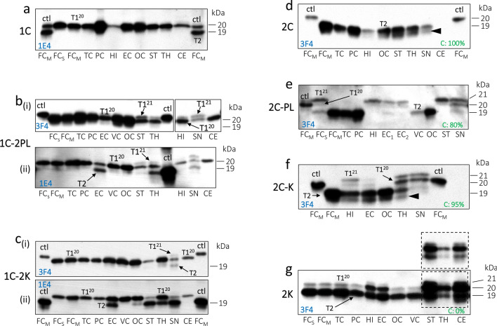
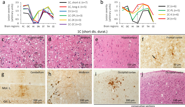
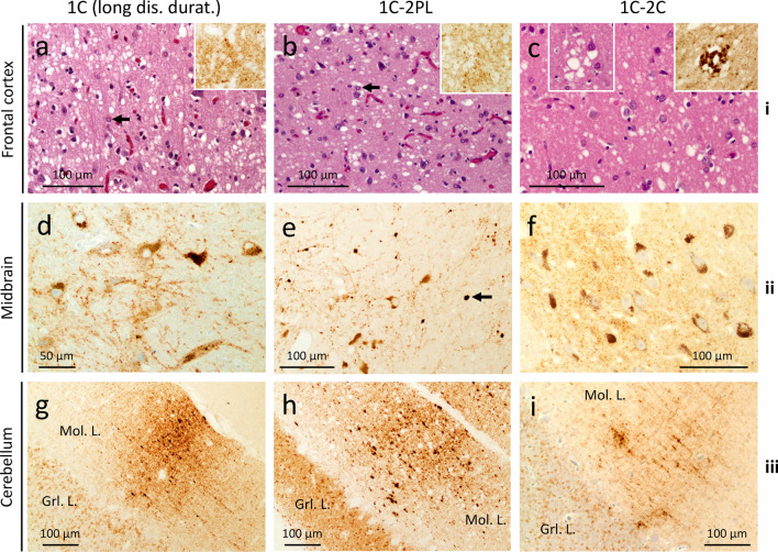
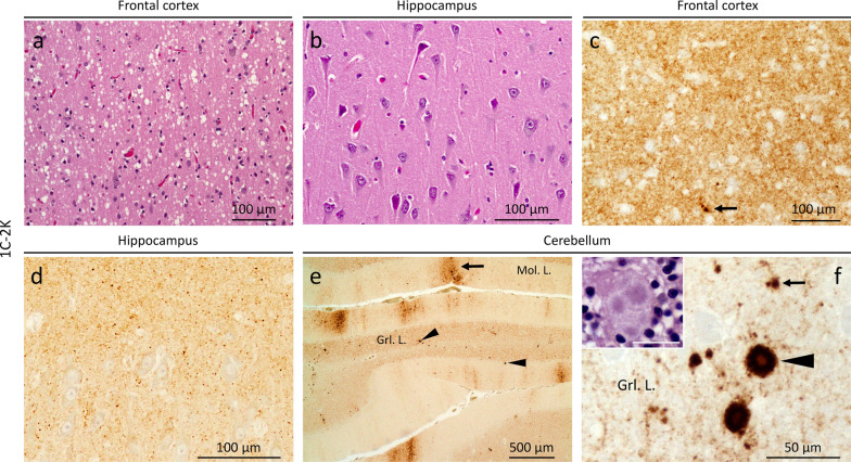
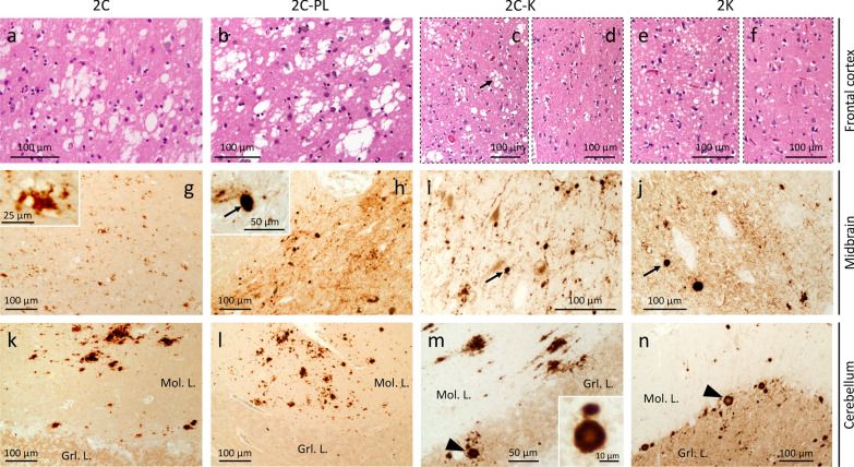
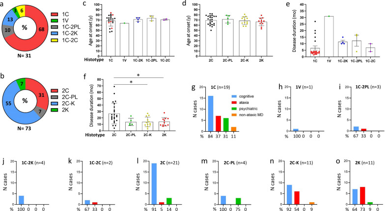
The MV1 and MV2 subtypes of sporadic Creutzfeldt-Jakob disease (sCJD) are linked to the heterozygous methionine (M)/valine (V) polymorphism at codon 129 of the prion protein (PrP) gene. MV2 is phenotypically heterogeneous, whereas MV1, due to its low prevalence, is one of the least well characterized subtypes. In this study, we investigated the biochemical properties of PrP and phenotypic expression of cases diagnosed as sCJD MV1 and MV2. We describe four MV2 histotypes: 2C, with cortical (C) coarse pathology; 2K, with kuru (K) plaque deposits; 2C-K, with co-existing C and K histotypic features; and the novel histotype 2C-PL that mimics 2C in the cerebral cortex and cerebellum, but exhibits plaque-like (PL) PrP deposits in subcortical regions (e.g., basal nuclei, thalamus and midbrain). Histotype prevalence is highest for 2C-K (55%), intermediate for 2C (31%), and lowest for 2C-PL and 2K (7%). Nearly every MV2 case expressed both PrP types, with T2 being the predominant type ("MV2-1"). MV1 cases typically show a rapid disease course (≤ 4 months), and feature the 1C histotype, phenotypically identical to sCJDMM1. Co-existing PrP types, with T1 significantly exceeding T2 ("MV1-2"), are detected in patients diagnosed as MV1 with longer disease courses. We observed four histotypes among MV1-2 cases, including two novel histotypes: 1V, reminiscent of sCJDVV1; 1C-2C, resembling sCJDMM1-2 with predominant MM1 histotypic component; and novel histotypes 1C-2PL and 1C-2K, overall mimicking 1C in the cerebral cortex, but harboring T2 and plaque-like PrP deposits in subcortical regions (1C-2PL), and T2 and kuru plaques in the cerebellum (1C-2K). Lesion profiles of 1C, 1V, and 1C-2C are similar, but differ from 1C-2PL and 1C-2K, as the latter two groups show prominent hippocampal and nigral degeneration. We believe that the novel "C-PL" histotypes are distinct entities rather than intermediate forms between "C" and "C-K" groups, and that 1C-2PL and 1C-2K histotypes may be characterized by different T1 variants of the same size.
MV1 和 MV2 亚型散发性克雅氏病(sCJD)与朊病毒蛋白(PrP)基因 129 位密码子的异亮氨酸/缬氨酸(M/V)多态性杂合有关。MV2 表型异质性,而 MV1 由于其患病率较低,是特征描述最少的亚型之一。在这项研究中,我们研究了被诊断为 sCJD MV1 和 MV2 的病例的 PrP 的生化特性和表型表达。我们描述了 4 种 MV2 组织型:2C,皮质(C)粗病理;2K,有库鲁(K)斑块沉积;2C-K,具有共存的 C 和 K 组织学特征;以及新型组织型 2C-PL,它在大脑皮层中模仿 2C,但在皮质下区域(例如基底核、丘脑和中脑)表现出斑块样(PL)PrP 沉积。2C-K 的组织型患病率最高(55%),2C 为中等(31%),2C-PL 和 2K 最低(7%)。几乎每个 MV2 病例都表达了两种 PrP 类型,其中 T2 是主要类型(“MV2-1”)。MV1 病例通常表现为快速病程(≤4 个月),特征为 1C 组织型,表型与 sCJDMM1 相同。在被诊断为 MV1 且病程较长的患者中,检测到共存的 PrP 类型,T1 显著超过 T2(“MV1-2”)。在 MV1-2 病例中观察到 4 种组织型,包括两种新型组织型:1V,类似于 sCJDVV1;1C-2C,类似于 sCJDMM1-2,主要为 MM1 组织学成分;以及新型组织型 1C-2PL 和 1C-2K,在大脑皮层中总体上模仿 1C,但在皮质下区域(1C-2PL)存在 T2 和斑块样 PrP 沉积,在小脑(1C-2K)存在 T2 和库鲁斑块。1C、1V 和 1C-2C 的病变谱相似,但与 1C-2PL 和 1C-2K 不同,后两者组表现出明显的海马和黑质变性。我们认为新型“C-PL”组织型是不同的实体,而不是“C”和“C-K”组之间的中间形式,并且 1C-2PL 和 1C-2K 组织型可能具有不同的相同大小的 T1 变体。