Liu Kai, Wang Sulong, Yalikun Ainizier, Ren Peng, Yusufu Aihemaitijiang
Department of Trauma and Microreconstructive Surgery, The First Affiliated Hospital of Xinjiang Medical University, Urumqi, Xinjiang, China.
Front Physiol. 2023 Sep 8;14:1259567. doi: 10.3389/fphys.2023.1259567. eCollection 2023.
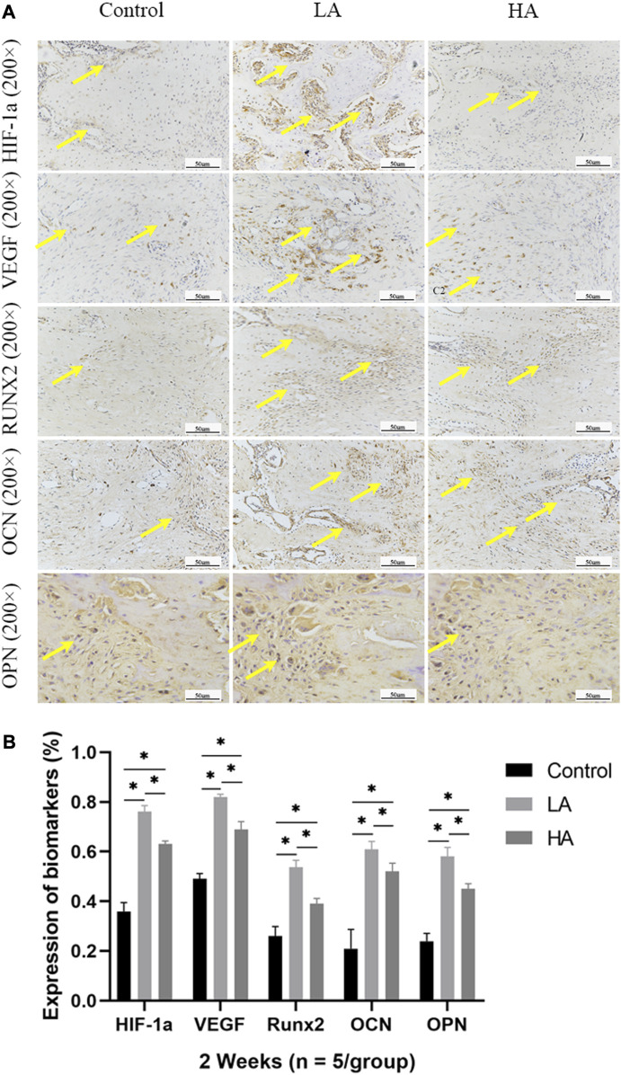
The purpose of this study was to observe the effect of the accordion technique (AT) during the distraction phase on chondrogenesis and bone regeneration in a rat femoral distraction osteogenesis (DO) model, and investigate its potential mechanism for reducing the total treatment time of DO. Fifty-four male Sprague-Dawley (SD) rats that were specific-pathogen-free (SPF) were subjected to DO surgery on the right femur. The distraction rate was 0.5 mm/day for 10 days, following a latency period of 5 days. Rats were randomly divided into Control (no AT, = 18), Group LA (low amplitude with AT, = 18), and Group HA (high amplitude with AT, = 18) according to different AT protocols in the distraction phase. Rats were respectively euthanized by anesthesia overdose at 2, 4 and 6 weeks of the consolidation phase, and the femurs were harvested. Digital radiography, micro-computed tomography (micro-CT), biomechanical tests, and histomorphological analysis were used to assess the quality of regenerated bone in the distraction area. Digital radiographic, micro-CT, biomechanical tests, and histological analysis revealed an increase in early-stage callus formation ( < 0.05) and improved blood supply to the callus tissue in Group LA, as compared to both the Control and Group HA. The enhanced differentiation of fibrous and cartilaginous tissue into bone tissue was also observed in Group LA, leading to improved strength and stiffness ( < 0.05) of the regenerated bone at 6 weeks of the consolidation phase. The angiogenic (hypoxia-inducible factor-1α (HIF-1α) and vascular endothelial growth factor (VEGF), < 0.05) and osteogenic (runt-related transcription factor 2 (RUNX2), osteocalcin (OCN) and osteopontin (OPN), < 0.05) biomarkers were higher expressed in Group LA at 2 and 4 weeks of consolidation phase, whereas decreased at 6 weeks of consolidation phase. The application of AT with low amplitude during the distraction phase can enhance chondrogenesis and bone regeneration by activating the angiogenesis factor pathway and upregulating the expression of osteogenic-related biomarkers such as HIF-1α, VEGF, RUNX2, OCN, and OPN.
本研究的目的是观察在大鼠股骨牵张成骨(DO)模型中,牵张期应用手风琴技术(AT)对软骨形成和骨再生的影响,并探讨其缩短DO总治疗时间的潜在机制。将54只无特定病原体(SPF)的雄性Sprague-Dawley(SD)大鼠右侧股骨进行DO手术。在5天的潜伏期后,以0.5毫米/天的速率牵张10天。根据牵张期不同的AT方案,将大鼠随机分为对照组(无AT,n = 18)、LA组(低幅度AT,n = 18)和HA组(高幅度AT,n = 18)。在巩固期的第2、4和6周,分别通过过量麻醉使大鼠安乐死,并采集股骨。采用数字X线摄影、微计算机断层扫描(micro-CT)、生物力学测试和组织形态学分析来评估牵张区域再生骨的质量。数字X线摄影、micro-CT、生物力学测试和组织学分析显示,与对照组和HA组相比,LA组早期骨痂形成增加(P < 0.05),骨痂组织血供改善。在LA组还观察到纤维和软骨组织向骨组织的分化增强,导致巩固期6周时再生骨的强度和刚度提高(P < 0.05)。在巩固期第2和4周,LA组血管生成(缺氧诱导因子-1α(HIF-1α)和血管内皮生长因子(VEGF),P < 0.05)和成骨( runt相关转录因子2(RUNX2)、骨钙素(OCN)和骨桥蛋白(OPN),P < 0.05)生物标志物表达较高,而在巩固期第6周降低。牵张期应用低幅度AT可通过激活血管生成因子途径并上调HIF-1α、VEGF、RUNX2、OCN和OPN等成骨相关生物标志物的表达,增强软骨形成和骨再生。