• 文献检索
  • 文档翻译
  • 深度研究
  • 学术资讯
  • Suppr Zotero 插件Zotero 插件
  • 邀请有礼
  • 套餐&价格
  • 历史记录
应用&插件
Suppr Zotero 插件Zotero 插件浏览器插件Mac 客户端Windows 客户端微信小程序
定价
高级版会员购买积分包购买API积分包
服务
文献检索文档翻译深度研究API 文档MCP 服务
关于我们
关于 Suppr公司介绍联系我们用户协议隐私条款
关注我们

Suppr 超能文献

核心技术专利:CN118964589B侵权必究
粤ICP备2023148730 号-1Suppr @ 2026

文献检索

告别复杂PubMed语法,用中文像聊天一样搜索,搜遍4000万医学文献。AI智能推荐,让科研检索更轻松。

立即免费搜索

文件翻译

保留排版,准确专业,支持PDF/Word/PPT等文件格式,支持 12+语言互译。

免费翻译文档

深度研究

AI帮你快速写综述,25分钟生成高质量综述,智能提取关键信息,辅助科研写作。

立即免费体验

锥形束计算机断层扫描(CBCT)扫描中上颌窦偶然异常的患病率:一项影像学研究。

Prevalence of Incidental Maxillary Sinus Anomalies on CBCT Scans: A Radiographic Study.

作者信息

Ahmed Junaid, Gupta Aditya, Shenoy Nandita, Sujir Nanditha, Muralidharan Archana

机构信息

Department of Oral Medicine and Radiology, Manipal College of Dental Sciences Mangalore, Manipal Academy of Higher Education, Manipal 575001, Karnataka, India.

出版信息

Diagnostics (Basel). 2023 Sep 12;13(18):2918. doi: 10.3390/diagnostics13182918.

DOI:10.3390/diagnostics13182918
PMID:37761284
原文链接:https://pmc.ncbi.nlm.nih.gov/articles/PMC10528184/
Abstract

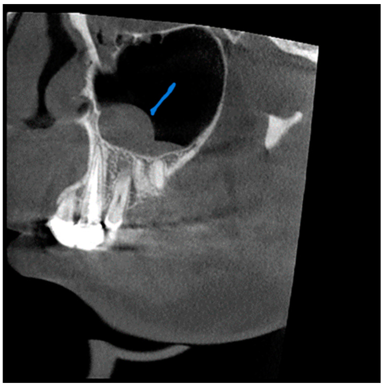
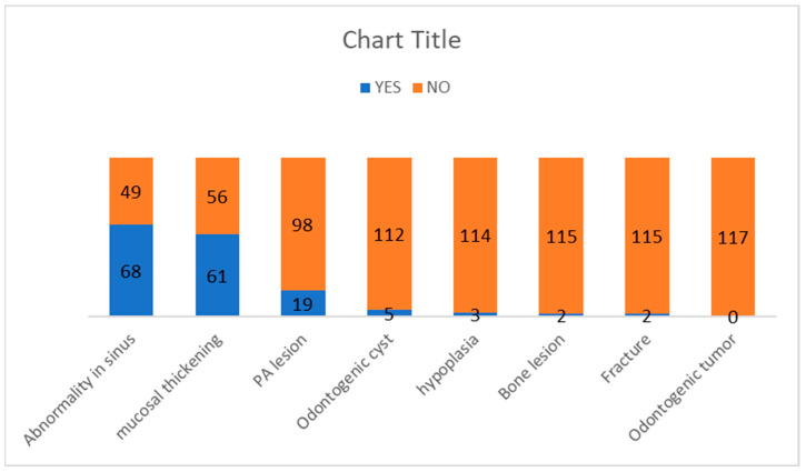
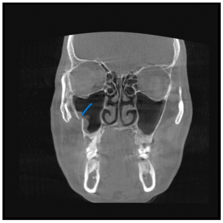
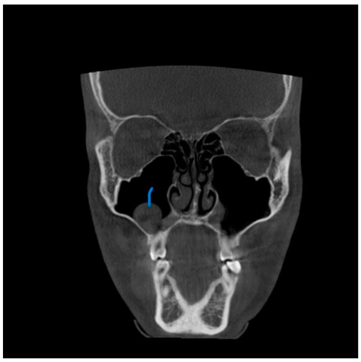
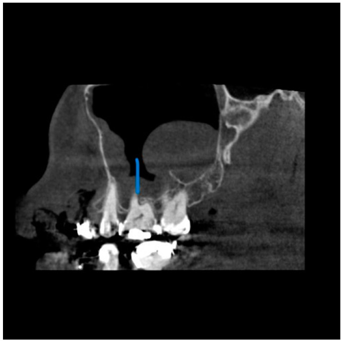
CBCT significantly impacts dental procedures and has brought significant reforms to our approach to diagnosis and treatment planning despite its limitations in differentiating soft tissues. It is an excellent imaging modality and quickly identifies sinus opacification and provides valuable insight into paranasal sinus pathologies, with considerably lower radiation exposure. The present study aimed to investigate the occurrence of maxillary sinus abnormalities in CBCT scans, identify the frequency, type, and location of these findings, and find the correlation between the distance of periapical lesions and radiographic changes in the maxillary sinus. Two examiners independently evaluated 117 patients to diagnose and classify the cases into different abnormality subtypes. The periapical lesions most closely related to the sinus were recorded. The diameters of the left and right maxillary sinus ostium and the distance of the ostium's lower border to the sinus's osseous floor were recorded. The findings were correlated with the age and gender of these patients. The present study reveals that sixty-one patients were diagnosed with mucosal thickening (52.1%). The sinus wall most affected by mucosal thickening was the maxillary sinus floor, followed by the medial and lateral walls. Of 19 patients with periapical lesions, 15 had maxillary sinus mucosal thickening, which is statistically significant ( = 0.004). The high occurrence of abnormalities in the maxillary sinus emphasizes the importance for the radiologist to comprehensively interpret the whole volume acquired in CBCT images, including the entire sinus. Incidental findings may be considered in the individual clinical context of signs and symptoms, reducing the risk of overestimating the real impact of radiographic findings.

摘要

尽管锥形束计算机断层扫描(CBCT)在区分软组织方面存在局限性,但它对牙科手术产生了重大影响,并给我们的诊断和治疗计划方法带来了重大变革。它是一种出色的成像方式,能快速识别鼻窦混浊,并为鼻旁窦病变提供有价值的见解,且辐射暴露量低得多。本研究旨在调查CBCT扫描中上颌窦异常的发生率,确定这些发现的频率、类型和位置,并找出根尖周病变距离与上颌窦影像学变化之间的相关性。两名检查人员独立评估了117例患者,以诊断并将病例分类为不同的异常亚型。记录与鼻窦关系最密切的根尖周病变。记录左右上颌窦口的直径以及窦口下缘至鼻窦骨底的距离。将这些发现与这些患者的年龄和性别相关联。本研究表明,61例患者被诊断为黏膜增厚(52.1%)。受黏膜增厚影响最大的鼻窦壁是上颌窦底,其次是内侧壁和外侧壁。在19例有根尖周病变的患者中,15例有上颌窦黏膜增厚,这具有统计学意义(P = 0.004)。上颌窦异常的高发生率强调了放射科医生全面解读CBCT图像中获取的整个容积(包括整个鼻窦)的重要性。偶然发现可在个体的症状和体征临床背景中予以考虑,以降低高估影像学发现实际影响的风险。

https://cdn.ncbi.nlm.nih.gov/pmc/blobs/9100/10528184/18e7111ca61a/diagnostics-13-02918-g005.jpg
https://cdn.ncbi.nlm.nih.gov/pmc/blobs/9100/10528184/b05bf2d0387e/diagnostics-13-02918-g001.jpg
https://cdn.ncbi.nlm.nih.gov/pmc/blobs/9100/10528184/82c09e24bc97/diagnostics-13-02918-g002.jpg
https://cdn.ncbi.nlm.nih.gov/pmc/blobs/9100/10528184/1540fcf97ab4/diagnostics-13-02918-g003.jpg
https://cdn.ncbi.nlm.nih.gov/pmc/blobs/9100/10528184/aef444a6d679/diagnostics-13-02918-g004.jpg
https://cdn.ncbi.nlm.nih.gov/pmc/blobs/9100/10528184/18e7111ca61a/diagnostics-13-02918-g005.jpg

相似文献

1
Prevalence of Incidental Maxillary Sinus Anomalies on CBCT Scans: A Radiographic Study.锥形束计算机断层扫描(CBCT)扫描中上颌窦偶然异常的患病率:一项影像学研究。
Diagnostics (Basel). 2023 Sep 12;13(18):2918. doi: 10.3390/diagnostics13182918.
2
Prevalence of Incidental Findings and Assessment of Maxillary Sinus Pathologies and Dental Diseases Using Cone-Beam Computed Tomography (CBCT) in the Tamil Nadu Population: A Retrospective Study.泰米尔纳德邦人群中使用锥形束计算机断层扫描(CBCT)对上颌窦病变和牙科疾病的偶然发现患病率及评估:一项回顾性研究
Cureus. 2024 Sep 8;16(9):e68929. doi: 10.7759/cureus.68929. eCollection 2024 Sep.
3
Prevalence of incidental maxillary sinus pathologies in dental patients on cone-beam computed tomographic images.牙科患者锥形束计算机断层扫描图像上上颌窦偶发病变的患病率。
Contemp Clin Dent. 2014 Jul;5(3):361-5. doi: 10.4103/0976-237X.137949.
4
Association of Inflammatory Periapical Lesions with Maxillary Sinus Abnormalities: a Retrospective Cone-Beam Computed Tomography Study.根尖周炎性病变与上颌窦异常的相关性:一项回顾性锥形束计算机断层扫描研究
J Dent (Shiraz). 2021 Dec;22(4):273-280. doi: 10.30476/DENTJODS.2021.87286.1254.
5
CBCT Evaluation of Periapical Pathologies in Maxillary Posterior Teeth and Their Relationship with Maxillary Sinus Mucosal Thickening.锥形束计算机断层扫描对上颌后牙根尖周病变及其与上颌窦黏膜增厚关系的评估
Healthcare (Basel). 2023 Mar 7;11(6):787. doi: 10.3390/healthcare11060787.
6
Evaluation of Association between Maxillary Posterior Teeth Periapical Pathologies and Maxillary Sinus Mucosal Changes-A Cone-Beam Computed Tomography (CBCT) Study.上颌后牙根尖周病变与上颌窦黏膜变化之间的相关性评估——一项锥形束计算机断层扫描(CBCT)研究
Indian J Radiol Imaging. 2023 Nov 23;34(2):246-253. doi: 10.1055/s-0043-1777013. eCollection 2024 Apr.
7
Occurrence of maxillary sinus abnormalities detected by cone beam CT in asymptomatic patients.无症状患者经锥形束 CT 检测出的上颌窦异常情况。
BMC Oral Health. 2012 Aug 10;12:30. doi: 10.1186/1472-6831-12-30.
8
Evaluation of Periapical Lesions and Their Association with Maxillary Sinus Abnormalities on Cone-beam Computed Tomographic Images.锥形束计算机断层扫描图像上根尖周病变及其与上颌窦异常的相关性评估
J Endod. 2016 Jan;42(1):42-6. doi: 10.1016/j.joen.2015.09.014. Epub 2015 Oct 29.
9
Incidence of Maxillary Sinus Disorders in Dental Patients Undergoing Cone-Beam Computed Tomography: A Retrospective Cross-Sectional Study.接受锥形束计算机断层扫描的牙科患者上颌窦疾病的发病率:一项回顾性横断面研究。
Cureus. 2024 Oct 8;16(10):e71114. doi: 10.7759/cureus.71114. eCollection 2024 Oct.
10
Incidental maxillary sinus findings in orthodontic patients: a radiographic analysis using cone-beam computed tomography (CBCT).正畸患者上颌窦偶然发现:锥形束 CT(CBCT)的放射分析。
Orthod Craniofac Res. 2011 Feb;14(1):17-24. doi: 10.1111/j.1601-6343.2010.01502.x. Epub 2010 Nov 22.

引用本文的文献

1
Evaluation of the relationship between periodontal bone loss and Schneiderian membrane thickness in upper posterior teeth with root tips associated with the maxillary sinus: a retrospective cone beam computed tomography study.根尖与上颌窦相关的上颌后牙牙周骨丧失与施耐德膜厚度之间关系的评估:一项回顾性锥形束计算机断层扫描研究
BMC Oral Health. 2025 Aug 20;25(1):1340. doi: 10.1186/s12903-025-06684-x.
2
Incidence of Maxillary Sinus Disorders in Dental Patients Undergoing Cone-Beam Computed Tomography: A Retrospective Cross-Sectional Study.接受锥形束计算机断层扫描的牙科患者上颌窦疾病的发病率:一项回顾性横断面研究。
Cureus. 2024 Oct 8;16(10):e71114. doi: 10.7759/cureus.71114. eCollection 2024 Oct.

本文引用的文献

1
Healing of Unilateral Maxillary Sinusitis by Endodontic and Periodontal Treatment of Maxillary Teeth.上颌牙的牙髓牙周联合治疗治愈单侧上颌窦炎。
Medicina (Kaunas). 2022 Sep 18;58(9):1302. doi: 10.3390/medicina58091302.
2
Evaluation of the effect of periapical lesions and other odontogenic conditions on maxillary sinus mucosal thickness characteristics and mucosal appearance: A CBCT study.根尖周病变及其他牙源性疾病对上颌窦黏膜厚度特征及黏膜外观影响的评估:一项锥形束计算机断层扫描(CBCT)研究
J Dent Res Dent Clin Dent Prospects. 2021 Summer;15(3):163-171. doi: 10.34172/joddd.2021.028. Epub 2021 Aug 25.
3
Association between Cone-Beam Computed Tomography and Histological and Immunohistochemical Features in Periapical Lesions Correlated with Thickened Maxillary Sinus Mucosa.
上颌窦黏膜增厚相关根尖周病变的锥形束 CT 与组织学及免疫组织化学特征的相关性研究。
Medicina (Kaunas). 2021 Aug 19;57(8):840. doi: 10.3390/medicina57080840.
4
Incidental findings in maxillary sinus area on cone-beam-computed-tomographic-scans: A retrospective study with emphasis on gender and ethnicity.锥形束计算机断层扫描在上颌窦区域的偶然发现:一项侧重于性别和种族的回顾性研究。
Saudi Dent J. 2021 May;33(4):184-187. doi: 10.1016/j.sdentj.2020.03.002. Epub 2020 Mar 18.
5
Quantitative analysis of metal artefacts of dental implant in CBCT image by correlation analysis to micro-CT: A microstructural study.通过与微 CT 的相关分析对 CBCT 图像中牙科种植体金属伪影的定量分析:一项结构研究。
Dentomaxillofac Radiol. 2021 Mar 1;50(3):20200365. doi: 10.1259/dmfr.20200365. Epub 2020 Oct 8.
6
A systematic review on incidental findings in cone beam computed tomography (CBCT) scans.锥形束计算机断层扫描(CBCT)扫描中的偶然发现的系统评价。
Dentomaxillofac Radiol. 2019 Oct;48(7):20180396. doi: 10.1259/dmfr.20180396. Epub 2019 Jun 28.
7
Three-dimensional evaluation of maxillary sinus volume in different age and sex groups using CBCT.使用锥形束计算机断层扫描(CBCT)对不同年龄和性别组的上颌窦容积进行三维评估。
Eur Arch Otorhinolaryngol. 2019 May;276(5):1493-1499. doi: 10.1007/s00405-019-05383-y. Epub 2019 Mar 16.
8
Incidental findings on cone-beam computed tomographic images: paranasal sinus findings and nasal septum variations.锥形束计算机断层扫描图像上的偶然发现:鼻窦发现及鼻中隔变异
Oral Radiol. 2018 Jan;34(1):40-48. doi: 10.1007/s11282-017-0283-y. Epub 2017 Mar 27.
9
Odontogenic sinusitis: A review of the current literature.牙源性鼻窦炎:当前文献综述
Laryngoscope Investig Otolaryngol. 2018 Mar 25;3(2):110-114. doi: 10.1002/lio2.147. eCollection 2018 Apr.
10
When Maxillary Sinusitis Does Not Heal: Findings on CBCT Scans of the Sinuses With a Particular Focus on the Occurrence of Odontogenic Causes of Maxillary Sinusitis.当上颌窦炎不愈合时:鼻窦CBCT扫描结果,特别关注上颌窦炎牙源性病因的发生情况。
Laryngoscope Investig Otolaryngol. 2017 Dec 11;2(6):442-446. doi: 10.1002/lio2.130. eCollection 2017 Dec.