de Alba Alvarado Mariana C, Torres Gutiérrez Elia, Cabrera Bravo Margarita, Zenteno Galindo Edgar, Villarreal Muñoz José Antonio, Salazar Schettino Paz María, Bucio Torres Martha Irene
Departamento de Microbiología y Parasitología, Facultad de Medicina, Universidad Nacional Autónoma de México, Coyoacán, Mexico City 04510, Mexico.
Departamento de Bioquímica, Facultad de Medicina, Universidad Nacional Autónoma de México, Coyoacán, Mexico City 04510, Mexico.
Pathogens. 2023 Aug 26;12(9):1084. doi: 10.3390/pathogens12091084.
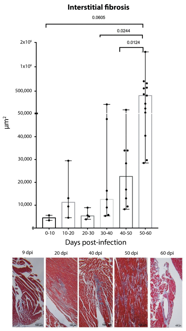
Symptoms in the acute phase of Chagas disease are usually mild and nonspecific. However, after several years, severe complications like dilated heart failure and even death may arise in the chronic phase. Due to the lack of specific symptoms in the acute phase, the aim of this work was to describe and analyze the cardiac histopathology during this phase in a CD1 mouse model by assessing parasitism, fibrotic damage, and the presence and composition of a cellular infiltrate, to determine its involvement in the pathogenesis of lesions in the cardiac tissue. Our results indicate that the acute phase lasts about 62 days post-infection (dpi). A significant increase in parasitemia was observed since 15 dpi, reaching a maximum at 33 dpi (4.1 × 10). The presence of amastigote nests was observed at 15-62 dpi, with a maximum count of 27 nests at 35 dpi. An infiltrate consisting primarily of macrophages and neutrophils was found in the cardiac tissue within the first 30 days, but the abundance of lymphocytes showed an 8 ≥ fold increase at 40-62 dpi. Unifocal interstitial fibrosis was identified after 9 dpi, which subsequently showed a 16 ≥ fold increase at 40-60 dpi, along with a 50% mortality rate in the model under study. The increased area of fibrotic lesions revealed progression in the extent of fibrosis, mainly at 50-62 dpi. The presence of perivasculitis and thrombus circulation disorders was seen in the last days (62 dpi); finally, cases of myocytolysis were observed at 50 and 62 dpi. These histopathological alterations, combined with collagen deposition, seem to lead to the development of interstitial fibrosis and damage to the cardiac tissue during the acute phase of infection. This study provides a more complete understanding of the patterns of histopathological abnormalities involved in the acute phase, which could help the development of new therapies to aid the preclinical tests of drugs for their application in Chagas disease.
恰加斯病急性期的症状通常较轻且无特异性。然而,数年后,慢性期可能会出现严重并发症,如扩张型心力衰竭甚至死亡。由于急性期缺乏特异性症状,本研究的目的是通过评估寄生虫感染、纤维化损伤以及细胞浸润的存在和组成,描述和分析CD1小鼠模型在该阶段的心脏组织病理学,以确定其在心脏组织病变发病机制中的作用。我们的结果表明,急性期持续约62天(感染后天数,dpi)。自感染后15 dpi起观察到寄生虫血症显著增加,在33 dpi时达到峰值(4.1×10)。在15 - 62 dpi观察到无鞭毛体巢的存在,在35 dpi时计数最高达27个巢。在最初30天内在心脏组织中发现主要由巨噬细胞和中性粒细胞组成的浸润,但在40 - 62 dpi时淋巴细胞数量增加了8倍以上。在感染后9 dpi确定存在单灶性间质纤维化,随后在40 - 60 dpi时增加了16倍以上,同时所研究模型的死亡率为50%。纤维化病变面积增加表明纤维化程度进展,主要发生在50 - 62 dpi。在最后几天(62 dpi)观察到血管周围炎和血栓循环障碍的存在;最后,在50和62 dpi观察到心肌溶解病例。这些组织病理学改变与胶原蛋白沉积相结合,似乎导致感染急性期间质纤维化的发展和心脏组织的损伤。本研究提供了对急性期所涉及的组织病理学异常模式更全面的理解,这有助于开发新的治疗方法,以辅助用于恰加斯病的药物的临床前测试。