• 文献检索
  • 文档翻译
  • 深度研究
  • 学术资讯
  • Suppr Zotero 插件Zotero 插件
  • 邀请有礼
  • 套餐&价格
  • 历史记录
应用&插件
Suppr Zotero 插件Zotero 插件浏览器插件Mac 客户端Windows 客户端微信小程序
定价
高级版会员购买积分包购买API积分包
服务
文献检索文档翻译深度研究API 文档MCP 服务
关于我们
关于 Suppr公司介绍联系我们用户协议隐私条款
关注我们

Suppr 超能文献

核心技术专利:CN118964589B侵权必究
粤ICP备2023148730 号-1Suppr @ 2026

文献检索

告别复杂PubMed语法,用中文像聊天一样搜索,搜遍4000万医学文献。AI智能推荐,让科研检索更轻松。

立即免费搜索

文件翻译

保留排版,准确专业,支持PDF/Word/PPT等文件格式,支持 12+语言互译。

免费翻译文档

深度研究

AI帮你快速写综述,25分钟生成高质量综述,智能提取关键信息,辅助科研写作。

立即免费体验

皮肤镜与反射式共聚焦显微镜在生殖器硬化性苔藓靶向治疗中的应用与比较:一项单臂前瞻性研究

Application and Comparison of Dermoscopy and Reflectance Confocal Microscopy in the Target Treatment of Genital Lichen Sclerosus: A Single-Arm Prospective Study.

作者信息

Bao Chengbei, Zhao Yan, Luo Renwei, Xu Qiuyun, Tong Zequn, Xiao Zhixun, Zhuang Zheyu, Dai Wenjia, Gu Bohan, Gong Ting, Cheng Bo, Ji Chao

机构信息

Department of Dermatology, The First Affiliated Hospital of Fujian Medical University, Fuzhou, 350000, Fujian, China.

Key Laboratory of Skin Cancer of Fujian Higher Education Institutions, The First Affiliated Hospital, Fujian Medical University, Fuzhou, 350000, Fujian, China.

出版信息

Dermatol Ther (Heidelb). 2023 Dec;13(12):3071-3084. doi: 10.1007/s13555-023-01039-2. Epub 2023 Oct 15.

DOI:10.1007/s13555-023-01039-2
PMID:37840118
原文链接:https://pmc.ncbi.nlm.nih.gov/articles/PMC10689603/
Abstract

INTRODUCTION

The treatment of genital lichen sclerosus (GLS) remains challenging. Baricitinib has been introduced in the treatment of GLS, but there's no imaging evaluation for GLS patients treated with it. No comparison of dermoscopy and reflectance confocal microscopy (RCM) assessments in GLS has been conducted. We performed this study to evaluate the efficacy and safety of baricitinib for GLS and to compare the value of dermoscopy and RCM assessments in GLS.

METHODS

Participants were treated with baricitinib for 6 months and assessed at week 0, 2, 4, 6, 8, and every 4 weeks for the next 16 weeks. All patients were evaluated for clinical, dermoscopic, and RCM variables, with numeric scores assigned to each parameter.

RESULTS

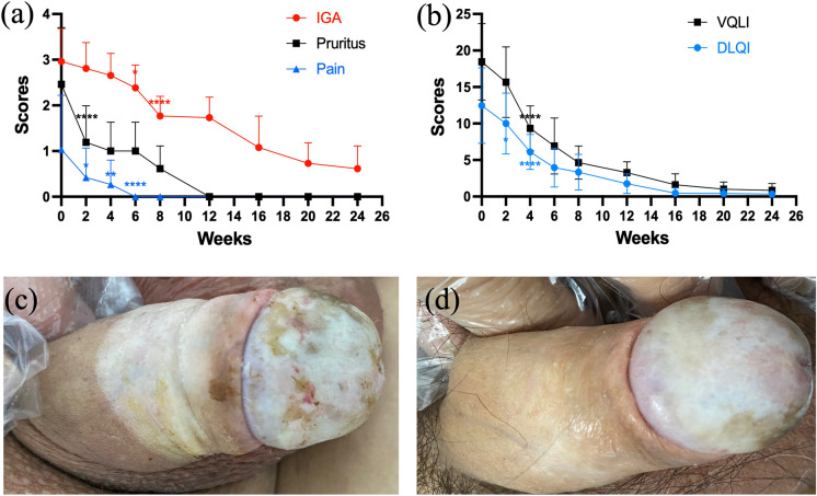
Twenty-six GLS patients were included in this study. All patients achieved Investigator's Global Assessment score ≤ 1 (with ≥ 2-grade improvement) at week 20. The scores of pruritus and pain decreased since week 2 (both P < 0.05). The DLQI and VQLI scores significantly decreased since week 4 (both P < 0.0001). White structureless areas improved at week 2 and white shiny streaks and follicular plugs improved at week 4 under dermoscopic examination. Vessels (P < 0.001) and brown structureless areas (P = 0.003) increased at week 8. In RCM, inflammatory cells count significantly decreased at week 2 (100.03 ± 33.24, P < 0.0001), with substantial regression at week 8 (16.98 ± 5.54, P < 0.0001). Epidermal thickness increased at week 12 (157.44 ± 37.87 μm versus 134.13 ± 36.60 μm, P = 0.0284). Irregular papillae, spongiosis, and fiber structures improved at week 20, week 4, and week 6 (all P < 0.01). Transient hypercholesterolemia (11.54%), thrombocytosis (7.69%), and elevated alanine aminotransferase (7.69%) occurred during treatment.

CONCLUSION

Both dermoscopy and RCM can be useful and non-invasive adjuvant tools for the evaluation and therapeutic monitoring of GLS. We recommended white structureless areas under dermoscopy and inflammatory cells count under RCM as variables for dermatologic imaging evaluation for GLS. Baricitinib is effective and safe for GLS, while randomized controlled trials are warranted.

摘要

引言

生殖器硬化性苔藓(GLS)的治疗仍然具有挑战性。巴瑞替尼已被用于GLS的治疗,但尚未对接受该药物治疗的GLS患者进行影像学评估。尚未对GLS的皮肤镜检查和反射式共聚焦显微镜(RCM)评估进行比较。我们开展这项研究以评估巴瑞替尼治疗GLS的疗效和安全性,并比较皮肤镜检查和RCM评估在GLS中的价值。

方法

参与者接受巴瑞替尼治疗6个月,并在第0、2、4、6、8周进行评估,之后的16周每4周评估一次。所有患者均接受临床、皮肤镜和RCM变量评估,每个参数都赋予了数值评分。

结果

本研究纳入了26例GLS患者。所有患者在第20周时达到研究者整体评估评分≤1(改善≥2级)。瘙痒和疼痛评分自第2周起下降(均P<0.05)。皮肤病生活质量指数(DLQI)和视觉功能生活质量指数(VQLI)评分自第4周起显著下降(均P<0.0001)。皮肤镜检查显示,白色无结构区域在第2周改善,白色发亮条纹和毛囊栓在第4周改善。血管(P<0.001)和棕色无结构区域(P=0.003)在第8周增加。在RCM中炎症细胞计数在第2周显著下降(100.03±33.24,P<0.0001),在第8周大幅消退(16.98±5.54,P<0.0001)。表皮厚度在第12周增加(157.44±37.87μm对134.13±36.60μm,P=0.0284)。不规则乳头、海绵形成和纤维结构在第20周、第4周和第6周改善(均P<0.01)。治疗期间出现短暂性高胆固醇血症(11.54%)、血小板增多症(7.69%)和丙氨酸转氨酶升高(7.69%)。

结论

皮肤镜检查和RCM均可作为评估和治疗监测GLS的有用且非侵入性的辅助工具。我们推荐将皮肤镜检查下的白色无结构区域和RCM下的炎症细胞计数作为GLS皮肤影像学评估的变量。巴瑞替尼治疗GLS有效且安全,但仍需进行随机对照试验。

https://cdn.ncbi.nlm.nih.gov/pmc/blobs/b06a/10689603/f00dfb1e24ba/13555_2023_1039_Fig4_HTML.jpg
https://cdn.ncbi.nlm.nih.gov/pmc/blobs/b06a/10689603/53911b362d32/13555_2023_1039_Fig1_HTML.jpg
https://cdn.ncbi.nlm.nih.gov/pmc/blobs/b06a/10689603/3aedf636b597/13555_2023_1039_Fig2_HTML.jpg
https://cdn.ncbi.nlm.nih.gov/pmc/blobs/b06a/10689603/58952ce6d6f1/13555_2023_1039_Fig3_HTML.jpg
https://cdn.ncbi.nlm.nih.gov/pmc/blobs/b06a/10689603/f00dfb1e24ba/13555_2023_1039_Fig4_HTML.jpg

相似文献

1
Application and Comparison of Dermoscopy and Reflectance Confocal Microscopy in the Target Treatment of Genital Lichen Sclerosus: A Single-Arm Prospective Study.皮肤镜与反射式共聚焦显微镜在生殖器硬化性苔藓靶向治疗中的应用与比较:一项单臂前瞻性研究
Dermatol Ther (Heidelb). 2023 Dec;13(12):3071-3084. doi: 10.1007/s13555-023-01039-2. Epub 2023 Oct 15.
2
Clinical and Dermoscopic Assessment of Vulvar Lichen Sclerosus After 5-Aminolevulinic Acid Photodynamic Therapy:A Prospective Study.5-氨基酮戊酸光动力疗法治疗外阴硬化性苔藓后的临床及皮肤镜评估:一项前瞻性研究
Photodiagnosis Photodyn Ther. 2021 Mar;33:102109. doi: 10.1016/j.pdpdt.2020.102109. Epub 2020 Nov 24.
3
Role of In Vivo Reflectance Confocal Microscopy in the Analysis of Melanocytic Lesions.体内反射共聚焦显微镜在黑素细胞性病变分析中的作用
Acta Dermatovenerol Croat. 2018 Apr;26(1):64-67.
4
Lentigo maligna and lentigo maligna melanoma in vivo differentiation with dermoscopy and reflectance confocal microscopy: A retrospective, multicentre study.利用皮肤镜和反射共聚焦显微镜对原位性恶性黑素细胞痣和恶性黑素瘤进行体内鉴别:一项回顾性、多中心研究。
J Eur Acad Dermatol Venereol. 2023 Nov;37(11):2293-2300. doi: 10.1111/jdv.19291. Epub 2023 Jul 21.
5
Comparison of dermoscopy and reflectance confocal microscopy accuracy for the diagnosis of psoriasis and lichen planus.比较皮肤镜检查和反射共聚焦显微镜对银屑病和扁平苔藓的诊断准确性。
Skin Res Technol. 2022 May;28(3):480-486. doi: 10.1111/srt.13158. Epub 2022 Mar 28.
6
Effects of topical piroxicam and sun filters in actinic keratosis evolution and field cancerization: a two-center, assessor-blinded, clinical, confocal microscopy and dermoscopy evaluation trial.吡罗昔康和防晒剂对光化性角化病进展和癌前病变的影响:一项双中心、评估者盲法、临床、共聚焦显微镜和皮肤镜评估试验。
Curr Med Res Opin. 2019 Oct;35(10):1785-1792. doi: 10.1080/03007995.2019.1626227. Epub 2019 Jun 13.
7
Assessing Light and Energy-Based Therapy by Optical Coherence Tomography and Reflectance Confocal Microscopy: A Randomized Trial of Photoaged Skin.通过光学相干断层扫描和反射式共聚焦显微镜评估基于光和能量的疗法:光老化皮肤的随机试验
Dermatology. 2022;238(3):422-429. doi: 10.1159/000517960. Epub 2021 Aug 27.
8
Amelanotic/hypomelanotic lentigo maligna: Dermoscopic and confocal features predicting diagnosis.无色素性/低色素性恶性雀斑样痣:预测诊断的皮肤镜和共聚焦特征。
J Eur Acad Dermatol Venereol. 2023 Feb;37(2):303-310. doi: 10.1111/jdv.18636. Epub 2022 Oct 17.
9
Melanocytic lesions with peripheral globules: results of an observational prospective study in 154 high-risk melanoma patients under digital dermoscopy follow-up evaluated with reflectance confocal microscopy.伴有周边小球的黑素细胞性病变:一项对154例高危黑色素瘤患者进行数字皮肤镜随访并采用反射式共聚焦显微镜评估的前瞻性观察研究结果
J Eur Acad Dermatol Venereol. 2021 May;35(5):1133-1142. doi: 10.1111/jdv.17105. Epub 2021 Feb 4.
10
Correlation of dermoscopic structures of melanocytic lesions to reflectance confocal microscopy.黑素细胞性皮损的皮肤镜结构与反射式共聚焦显微镜检查的相关性
Arch Dermatol. 2007 Feb;143(2):176-85. doi: 10.1001/archderm.143.2.176.

引用本文的文献

1
Utilizing reflectance confocal microscopy for surveillance of neoplastic changes in penile lichen sclerosus: A report of two cases.利用反射式共聚焦显微镜监测阴茎硬化性苔藓的肿瘤性变化:两例报告
JAAD Case Rep. 2024 Jun 5;50:59-61. doi: 10.1016/j.jdcr.2024.05.028. eCollection 2024 Aug.

本文引用的文献

1
Abrocitinib as a novel treatment for lichen sclerosus.阿布昔替尼作为一种治疗硬化性苔藓的新型疗法。
Br J Dermatol. 2023 Jul 7;189(1):136-138. doi: 10.1093/bjd/ljad129.
2
Mid-term symptomatic relief after platelet-rich plasma infiltration in vulvar lichen sclerosus.富血小板血浆浸润治疗外阴硬化性苔藓的中期症状缓解。
Arch Dermatol Res. 2023 Aug;315(6):1527-1532. doi: 10.1007/s00403-023-02529-1. Epub 2023 Jan 19.
3
[Inflammatory dermatoses in skin of color].[有色人种皮肤的炎症性皮肤病]
Dermatologie (Heidelb). 2023 Feb;74(2):84-89. doi: 10.1007/s00105-022-05096-0. Epub 2023 Jan 2.
4
Reflectance confocal microscopy for non-invasive diagnosis of childhood-onset vulvar lichen sclerosus: A retrospective study.反射式共聚焦显微镜用于儿童期外阴硬化性苔藓的无创诊断:一项回顾性研究。
J Eur Acad Dermatol Venereol. 2023 Mar;37(3):e303-e305. doi: 10.1111/jdv.18786. Epub 2022 Dec 7.
5
Baricitinib for the treatment of cutaneous dermatomyositis: A prospective, open-label study.巴瑞替尼治疗皮肤型皮肌炎:一项前瞻性、开放标签研究。
J Am Acad Dermatol. 2022 Dec;87(6):1374-1376. doi: 10.1016/j.jaad.2022.08.025. Epub 2022 Aug 20.
6
Oral tofacitinib for successful treatment of refractory alopecia areata in preschool children.口服托法替布成功治疗学龄前儿童难治性斑秃
J Eur Acad Dermatol Venereol. 2022 Dec;36(12):e1055-e1057. doi: 10.1111/jdv.18447. Epub 2022 Sep 22.
7
Successfully treated extragenital lichen sclerosus in a 2-year-old boy by baricitinib assessed by dermoscopy: A case report.
Dermatol Ther. 2022 Sep;35(9):e15712. doi: 10.1111/dth.15712. Epub 2022 Jul 19.
8
Exploring reflectance confocal microscopy as a non-invasive diagnostic tool for genital lichen sclerosus.探索反射式共聚焦显微镜作为一种用于诊断外阴硬化性苔藓的非侵入性工具。
Exp Ther Med. 2022 Jun;23(6):410. doi: 10.3892/etm.2022.11337. Epub 2022 Apr 26.
9
Influence of Photodynamic Therapy on Lichen Sclerosus with Neoplastic Background.光动力疗法对伴有肿瘤背景的硬化性苔藓的影响。
J Clin Med. 2022 Feb 19;11(4):1100. doi: 10.3390/jcm11041100.
10
Autoinflammatory Keratinization Disease With Hepatitis and Autism Reveals Roles for JAK1 Kinase Hyperactivity in Autoinflammation.自身炎症性角化病伴发肝炎和自闭症揭示 JAK1 激酶活性过度在自身炎症中的作用。
Front Immunol. 2022 Jan 3;12:737747. doi: 10.3389/fimmu.2021.737747. eCollection 2021.