Department of Obstetrics and Gynecology, University of Fukui, 23-3 Matsuoka-Shimoaizuki, Eiheiji-cho, Yoshida-gun, Fukui, 910-1193, Japan.
Department of Radiology, University of Fukui, Fukui, Japan.
Sci Rep. 2023 Nov 1;13(1):18864. doi: 10.1038/s41598-023-46261-2.
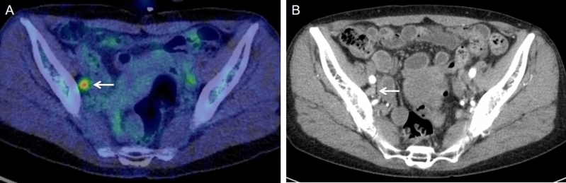
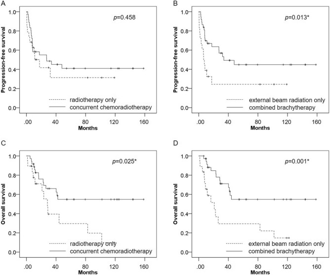
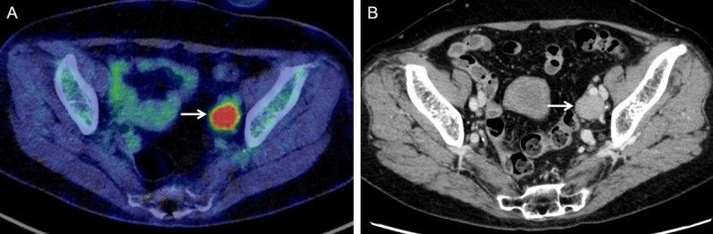
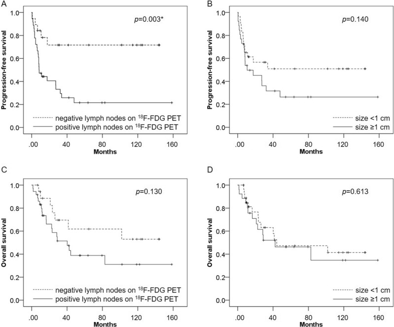
The effect on survival of radiographic lymph node metastasis in uterine cervical cancer patients is more important than before, even though its prognostic value not been well investigated. The aim of our study is to evaluate the prognostic potential of F-fluorodeoxyglucose Positron Emission Tomography (F-FDG PET) compared with Computed Tomography (CT) in uterine cervical cancer patients with stage IIICr allocated by imaging. Fifty-five patients with biopsy-proven primary cervical cancer underwent definitive radiation therapy for stages IIB-IVB of The International Federation of Gynecology and Obstetrics (FIGO) 2018 classifications. The prognostic performance of pretreatment F-FDG PET and CT for assessing lymph node metastasis was evaluated by two experienced readers. The PET and CT findings were correlated with the risk of progression-free survival (PFS) and overall survival (OS). Kaplan-Meier survival curves showed that PFS was significantly worse in patients with positive lymph nodes on F-FDG PET than in those patients with negative lymph nodes on F-FDG PET (p = 0.003), whereas there was no significant difference in PFS between patients with lymph nodes sized ≥ 1 cm and those sized < 1 cm (p = 0.140). Univariate analysis showed that positive lymph nodes on F-FDG PET was significantly associated with poor PFS (p = 0.006), whereas lymph node size was not significantly associated with poor PFS (p = 0.145). In multivariate analysis, positive lymph nodes on F-FDG PET was significantly associated with poor PFS (p = 0.006) and was an independent prognostic factor for PFS. F-FDG PET offers high prognostic value for patients with stage IIICr allocated by imaging compared with CT, suggesting that F-FDG PET might be useful in clinical staging decisions and thus promote optimal diagnostic and therapeutic strategies.
影像学分期为 IIICr 的宫颈癌患者,其生存受淋巴结转移的影响比以往更为重要,尽管其预后价值尚未得到充分研究。本研究旨在评估氟-18 氟代脱氧葡萄糖正电子发射断层扫描(F-FDG PET)与计算机断层扫描(CT)在影像学分期为 IIICr 的宫颈癌患者中的预后价值。55 例经活检证实的原发性宫颈癌患者接受了根治性放疗,分期为国际妇产科联合会(FIGO)2018 分类的 IIB-IVB 期。两名有经验的阅片者评估了治疗前 F-FDG PET 和 CT 评估淋巴结转移的预后性能。PET 和 CT 结果与无进展生存期(PFS)和总生存期(OS)的风险相关。Kaplan-Meier 生存曲线显示,F-FDG PET 阳性淋巴结患者的 PFS 明显差于 F-FDG PET 阴性淋巴结患者(p=0.003),而淋巴结大小≥1cm 和<1cm 的患者 PFS 无显著差异(p=0.140)。单因素分析显示,F-FDG PET 阳性淋巴结与较差的 PFS 显著相关(p=0.006),而淋巴结大小与较差的 PFS 无显著相关(p=0.145)。多因素分析显示,F-FDG PET 阳性淋巴结与较差的 PFS 显著相关(p=0.006),是 PFS 的独立预后因素。与 CT 相比,F-FDG PET 对影像学分期为 IIICr 的患者具有更高的预后价值,提示 F-FDG PET 可能有助于临床分期决策,从而促进最佳诊断和治疗策略。