• 文献检索
  • 文档翻译
  • 深度研究
  • 学术资讯
  • Suppr Zotero 插件Zotero 插件
  • 邀请有礼
  • 套餐&价格
  • 历史记录
应用&插件
Suppr Zotero 插件Zotero 插件浏览器插件Mac 客户端Windows 客户端微信小程序
定价
高级版会员购买积分包购买API积分包
服务
文献检索文档翻译深度研究API 文档MCP 服务
关于我们
关于 Suppr公司介绍联系我们用户协议隐私条款
关注我们

Suppr 超能文献

核心技术专利:CN118964589B侵权必究
粤ICP备2023148730 号-1Suppr @ 2026

文献检索

告别复杂PubMed语法,用中文像聊天一样搜索,搜遍4000万医学文献。AI智能推荐,让科研检索更轻松。

立即免费搜索

文件翻译

保留排版,准确专业,支持PDF/Word/PPT等文件格式,支持 12+语言互译。

免费翻译文档

深度研究

AI帮你快速写综述,25分钟生成高质量综述,智能提取关键信息,辅助科研写作。

立即免费体验

通过超声造影评估贝伐单抗化疗对结直肠癌肝转移的治疗反应

Treatment response assessment to chemotherapy with bevacizumab for colorectal liver metastasis by contrast-enhanced ultrasound.

作者信息

Wu Wen-Qing, Wang Xi, Dong Cai-Hong, Mao Li-Juan, Wang Han-Tao, Lu Qing

机构信息

Department of Ultrasound, Zhongshan Hospital, Fudan University, Shanghai, China.

Shanghai Institute of Medical Imaging, Fudan University, Shanghai, China.

出版信息

Quant Imaging Med Surg. 2024 Jan 3;14(1):548-565. doi: 10.21037/qims-23-1027. Epub 2023 Nov 7.

DOI:10.21037/qims-23-1027
PMID:38223071
原文链接:https://pmc.ncbi.nlm.nih.gov/articles/PMC10784026/
Abstract

BACKGROUND

Though contrast-enhanced ultrasound (CEUS) perfusion parameters have been approved to be potential indicators for response to chemotherapy in solid tumors, their ability in assessment of colorectal liver metastasis (CRLM) to chemotherapy with bevacizumab (Bev) has rarely been investigated.

METHODS

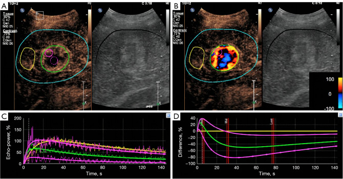
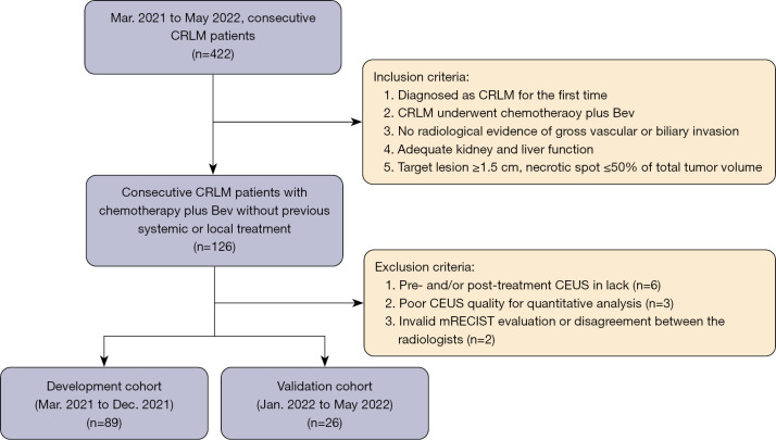
From March 2021 to May 2022, 115 consecutive CRLM patients with CEUS pre- and post-2 months' chemotherapy with Bev were prospectively enrolled. One target lesion per patient underwent CEUS quantitative analysis with SonoLiver software. Rise time, time-to-peak, mean transit time, maximal intensity (IMAX), and area under the time-intensity curve (AUC) were assessed with region of interest (ROI) selected on whole lesion, lesion periphery, and internal lesion, respectively. The reduction and ratio of post- to pre-treatment in parameters were investigated in development cohort (n=89) and validated in internal validation cohort (n=26) according to the chronological order.

RESULTS

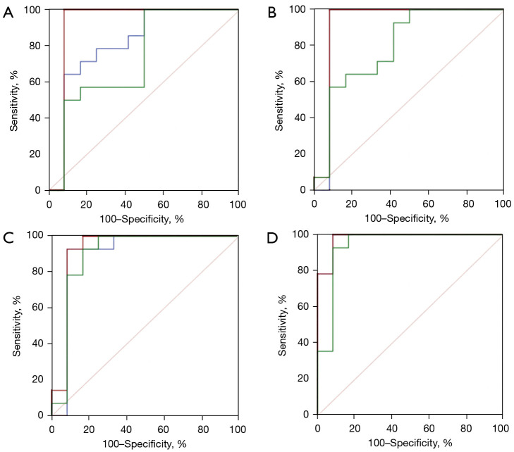
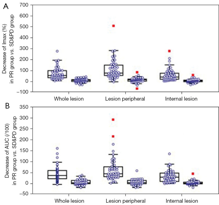
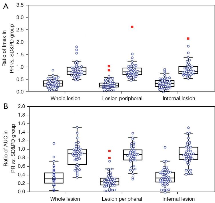
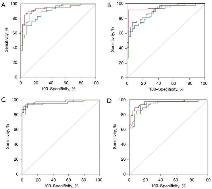
With modified Response Evaluation Criteria in Solid Tumor as reference, 48, 14 responders and 41, 12 non-responders were included in development and validation cohort, respectively. Significantly smaller values of IMAX and AUC on ROIwhole, ROIperipheral, and ROIinternal, were observed post-treatment in development cohort (all P<0.05). In predicting treatment response, the influence of ROI selection was observed when using ∆IMAX and ∆AUC, while no influence was observed using ratios. Areas under the receiver operating characteristic curve (AUROCs) for ∆IMAX and ∆AUC on ROIperipheral were 0.939 (0.867-0.979), 0.951 (0.883-0.985), and 0.917 (0.740-0.988), 0.923 (0.748-0.990) in development and validation cohort, respectively. For ratios of IMAX and AUC, AUROCs were 0.976 (0.919-0.997), 0.938 (0.865-0.978), and 0.899 (0.717-0.982), 0.982 (0.836-1.000) in development and validation cohort, respectively.

CONCLUSIONS

IMAX and AUC showed significant reductions in responders, and different analyses ROIs influence the performance of ∆IMAX and ∆AUC in response assessment. Parameters derived from ROI peripheral exhibited the most promising results in predicting treatment response.

摘要

背景

尽管超声造影(CEUS)灌注参数已被认可为实体瘤化疗反应的潜在指标,但它们在评估结直肠癌肝转移(CRLM)对贝伐单抗(Bev)化疗反应中的能力鲜有研究。

方法

2021年3月至2022年5月,前瞻性纳入115例连续的CRLM患者,这些患者在接受Bev化疗前和化疗2个月后均接受了CEUS检查。每位患者的一个靶病灶使用SonoLiver软件进行CEUS定量分析。分别在整个病灶、病灶周边和病灶内部选择感兴趣区(ROI),评估上升时间、达峰时间、平均通过时间、最大强度(IMAX)和时间-强度曲线下面积(AUC)。根据时间顺序,在开发队列(n=89)中研究参数治疗后较治疗前的降低值和比值,并在内部验证队列(n=26)中进行验证。

结果

以实体瘤改良反应评估标准为参照,开发队列和验证队列分别纳入48例、14例反应者和41例、12例无反应者。在开发队列中,治疗后观察到ROI整体、ROI周边和ROI内部的IMAX和AUC值显著更小(均P<0.05)。在预测治疗反应时,使用∆IMAX和∆AUC时观察到ROI选择的影响,而使用比值时未观察到影响。开发队列和验证队列中,ROI周边的∆IMAX和∆AUC的受试者工作特征曲线下面积(AUROC)分别为0.939(0.867-0.979)、0.951(0.883-0.985)和0.917(0.740-0.988)、0.923(0.748-0.990)。对于IMAX和AUC的比值,开发队列和验证队列的AUROC分别为0.976(0.919-0.997)、0.938(0.865-0.978)和0.899(0.717-0.982)、0.982(0.836-1.000)。

结论

反应者的IMAX和AUC显著降低,不同的分析ROI会影响∆IMAX和∆AUC在反应评估中的性能。来自ROI周边的参数在预测治疗反应方面显示出最有前景的结果。

https://cdn.ncbi.nlm.nih.gov/pmc/blobs/5542/10784026/5e99534ce126/qims-14-01-548-f6.jpg
https://cdn.ncbi.nlm.nih.gov/pmc/blobs/5542/10784026/a3004eae27de/qims-14-01-548-f1.jpg
https://cdn.ncbi.nlm.nih.gov/pmc/blobs/5542/10784026/16b8f57cedaf/qims-14-01-548-f2.jpg
https://cdn.ncbi.nlm.nih.gov/pmc/blobs/5542/10784026/04a48b108022/qims-14-01-548-f3.jpg
https://cdn.ncbi.nlm.nih.gov/pmc/blobs/5542/10784026/1534ace7f12f/qims-14-01-548-f4.jpg
https://cdn.ncbi.nlm.nih.gov/pmc/blobs/5542/10784026/940fb9b447bb/qims-14-01-548-f5.jpg
https://cdn.ncbi.nlm.nih.gov/pmc/blobs/5542/10784026/5e99534ce126/qims-14-01-548-f6.jpg

相似文献

1
Treatment response assessment to chemotherapy with bevacizumab for colorectal liver metastasis by contrast-enhanced ultrasound.通过超声造影评估贝伐单抗化疗对结直肠癌肝转移的治疗反应
Quant Imaging Med Surg. 2024 Jan 3;14(1):548-565. doi: 10.21037/qims-23-1027. Epub 2023 Nov 7.
2
Value of Perfusion Parameters for Differentiating Hepatocellular Carcinoma and Liver Metastasis With Hypervascularity and a Normal Hepatic Background on Contrast-Enhanced Ultrasound Imaging.超声造影增强成像背景正常肝实质中高血供肝细胞癌与肝转移瘤的灌注参数鉴别价值。
J Ultrasound Med. 2019 Oct;38(10):2601-2608. doi: 10.1002/jum.14957. Epub 2019 Jan 30.
3
Can quantitative contrast-enhanced ultrasonography predict cervical tumor response to neoadjuvant chemotherapy?定量对比增强超声检查能否预测宫颈癌新辅助化疗的疗效?
Eur J Radiol. 2016 Nov;85(11):2111-2118. doi: 10.1016/j.ejrad.2016.09.025. Epub 2016 Sep 25.
4
Quantitative analysis of pre-treatment dynamic contrast-enhanced ultrasound for assessing the response of colorectal liver metastases to chemotherapy plus targeted therapy: a dual-institutional study.治疗前动态对比增强超声定量分析评估结直肠癌肝转移化疗联合靶向治疗反应的双中心研究。
Abdom Radiol (NY). 2024 Feb;49(2):414-424. doi: 10.1007/s00261-023-04055-0. Epub 2023 Oct 18.
5
Quantitative Evaluation of Combretastatin A4 Phosphate Early Efficacy in a Tumor Model with Dynamic Contrast-Enhanced Ultrasound.在动态对比增强超声肿瘤模型中对磷酸考布他汀A4早期疗效的定量评估
Ultrasound Med Biol. 2018 Apr;44(4):840-852. doi: 10.1016/j.ultrasmedbio.2017.12.002. Epub 2018 Feb 1.
6
Quantitative Analysis of Perfusion Characteristics Using Contrast-Enhanced Ultrasound in Patients with Choroidal Metastasis.应用对比增强超声对脉络膜转移瘤患者灌注特征的定量分析。
Ophthalmic Res. 2022;65(1):86-93. doi: 10.1159/000510777. Epub 2020 Aug 11.
7
Regions of interest and parameters for the quantitative analysis of contrast-enhanced ultrasound to evaluate the anti-angiogenic effects of bevacizumab.感兴趣区域和参数的定量分析对比增强超声评估贝伐单抗的抗血管生成作用。
Mol Med Rep. 2013 Jul;8(1):154-60. doi: 10.3892/mmr.2013.1499. Epub 2013 May 29.
8
Prediction of treatment responses to neoadjuvant chemotherapy in breast cancer using contrast-enhanced ultrasound.使用超声造影预测乳腺癌新辅助化疗的治疗反应
Gland Surg. 2021 Apr;10(4):1280-1290. doi: 10.21037/gs-20-836.
9
Application of Quantitative Parameters of Contrast-Enhanced Ultrasound in Common Benign and Malignant Lesions in Pediatric Livers: A Preliminary Study.超声造影定量参数在小儿肝脏常见良恶性病变中的应用:一项初步研究
Diagnostics (Basel). 2023 Nov 14;13(22):3443. doi: 10.3390/diagnostics13223443.
10
Response evaluation of chemotherapy in metastatic colorectal cancer by contrast enhanced ultrasound.对比增强超声在转移性结直肠癌化疗中的疗效评价。
World J Gastroenterol. 2012 Feb 14;18(6):541-5. doi: 10.3748/wjg.v18.i6.541.

本文引用的文献

1
Quantitative analysis of contrast-enhanced ultrasonography in rat models of hepatic acute graft-versus-host disease.肝急性移植物抗宿主病大鼠模型中超声造影的定量分析
Quant Imaging Med Surg. 2023 Aug 1;13(8):4908-4918. doi: 10.21037/qims-22-1145. Epub 2023 May 22.
2
Evaluation of peripheral enhancement on contrast-enhanced computed tomography and corresponding pathological findings in colorectal liver metastases after preoperative chemotherapy.术前化疗后结直肠癌肝转移患者的对比增强计算机断层扫描外周强化评估及相应病理结果
Pol J Radiol. 2023 May 16;88:e251-e255. doi: 10.5114/pjr.2023.127611. eCollection 2023.
3
Comparison of superb microvascular imaging (SMI) quantified with ImageJ to quantified contrast-enhanced ultrasound (qCEUS) in liver metastases-a pilot study.
使用ImageJ量化的超微血管成像(SMI)与肝脏转移瘤中量化对比增强超声(qCEUS)的比较——一项初步研究
Quant Imaging Med Surg. 2022 Mar;12(3):1762-1774. doi: 10.21037/qims-21-383.
4
Enhanced Rim on MDCT of Colorectal Liver Metastases: Assessment of Ability to Predict Progression-Free Survival and Response to Bevacizumab-Based Chemotherapy.结直肠肝转移瘤 MDCT 增强环征:预测无进展生存期和贝伐珠单抗为基础化疗反应能力的评估。
AJR Am J Roentgenol. 2020 Dec;215(6):1377-1383. doi: 10.2214/AJR.19.22280. Epub 2020 Sep 29.
5
Early response evaluation during preoperative chemotherapy for colorectal liver metastases: Combined size and morphology-based criteria predict pathological response and survival after resection.结直肠癌肝转移术前化疗期间的早期反应评估:基于大小和形态的联合标准可预测切除术后的病理反应和生存情况。
J Surg Oncol. 2020 Feb;121(2):382-391. doi: 10.1002/jso.25796. Epub 2019 Dec 1.
6
Morphologic Response and Tumor Shrinkage as Early Predictive Markers in Unresectable Colorectal Liver Metastases.形态学反应和肿瘤缩小作为不可切除结直肠癌肝转移早期预测标志物
Anticancer Res. 2018 Nov;38(11):6501-6506. doi: 10.21873/anticanres.13014.
7
Correlation between CT morphologic appearance and histologic findings in colorectal liver metastasis after preoperative chemotherapy.结直肠癌肝转移患者化疗术前 CT 形态学表现与组织学发现的相关性。
Abdom Radiol (NY). 2018 Nov;43(11):2991-3000. doi: 10.1007/s00261-018-1588-y.
8
Comparison of early radiological predictors of outcome in patients with colorectal cancer with unresectable hepatic metastases treated with bevacizumab.比较贝伐珠单抗治疗不可切除肝转移结直肠癌患者的早期影像学预测因子与预后的关系。
Gut. 2018 Jun;67(6):1095-1102. doi: 10.1136/gutjnl-2017-313786. Epub 2017 Oct 30.
9
Early Recurrence After Hepatectomy for Colorectal Liver Metastases: What Optimal Definition and What Predictive Factors?结直肠癌肝转移肝切除术后的早期复发:最佳定义及预测因素是什么?
Oncologist. 2016 Jul;21(7):887-94. doi: 10.1634/theoncologist.2015-0468. Epub 2016 Apr 28.
10
Cancer statistics in China, 2015.《中国癌症统计数据 2015》
CA Cancer J Clin. 2016 Mar-Apr;66(2):115-32. doi: 10.3322/caac.21338. Epub 2016 Jan 25.