• 文献检索
  • 文档翻译
  • 深度研究
  • 学术资讯
  • Suppr Zotero 插件Zotero 插件
  • 邀请有礼
  • 套餐&价格
  • 历史记录
应用&插件
Suppr Zotero 插件Zotero 插件浏览器插件Mac 客户端Windows 客户端微信小程序
定价
高级版会员购买积分包购买API积分包
服务
文献检索文档翻译深度研究API 文档MCP 服务
关于我们
关于 Suppr公司介绍联系我们用户协议隐私条款
关注我们

Suppr 超能文献

核心技术专利:CN118964589B侵权必究
粤ICP备2023148730 号-1Suppr @ 2026

文献检索

告别复杂PubMed语法,用中文像聊天一样搜索,搜遍4000万医学文献。AI智能推荐,让科研检索更轻松。

立即免费搜索

文件翻译

保留排版,准确专业,支持PDF/Word/PPT等文件格式,支持 12+语言互译。

免费翻译文档

深度研究

AI帮你快速写综述,25分钟生成高质量综述,智能提取关键信息,辅助科研写作。

立即免费体验

用于脑水肿的新型先进成像技术。

Novel advanced imaging techniques for cerebral oedema.

作者信息

Pham Jenny, Ng Felix C

机构信息

Department of Radiology, Royal Melbourne Hospital, Parkville, VIC, Australia.

Department of Neurology, Royal Melbourne Hospital, Parkville, VIC, Australia.

出版信息

Front Neurol. 2024 Jan 31;15:1321424. doi: 10.3389/fneur.2024.1321424. eCollection 2024.

DOI:10.3389/fneur.2024.1321424
PMID:38356883
原文链接:https://pmc.ncbi.nlm.nih.gov/articles/PMC10865379/
Abstract

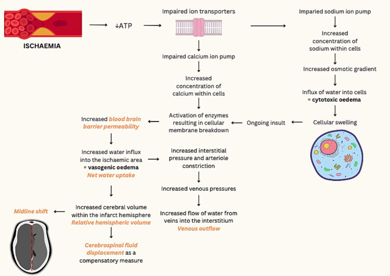
Cerebral oedema following acute ischemic infarction has been correlated with poor functional outcomes and is the driving mechanism of malignant infarction. Measurements of midline shift and qualitative assessment for herniation are currently the main CT indicators for cerebral oedema but have limited sensitivity for small cortical infarcts and are typically a delayed sign. In contrast, diffusion-weighted (DWI) or T2-weighted magnetic resonance imaging (MRI) are highly sensitive but are significantly less accessible. Due to the need for early quantification of cerebral oedema, several novel imaging biomarkers have been proposed. Based on neuroanatomical shift secondary to space-occupying oedema, measures such as relative hemispheric volume and cerebrospinal fluid displacement are correlated with poor outcomes. In contrast, other imaging biometrics, such as net water uptake, T2 relaxometry and blood brain barrier permeability, reflect intrinsic tissue changes from the influx of fluid into the ischemic region. This review aims to discuss quantification of cerebral oedema using current and developing advanced imaging techniques, and their role in predicting clinical outcomes.

摘要

急性缺血性梗死后脑水肿与功能预后不良相关,是恶性梗死的驱动机制。目前,中线移位测量和脑疝的定性评估是脑水肿的主要CT指标,但对小皮质梗死的敏感性有限,且通常是延迟征象。相比之下,扩散加权(DWI)或T2加权磁共振成像(MRI)高度敏感,但可及性显著较低。由于需要对脑水肿进行早期量化,已提出了几种新型成像生物标志物。基于占位性水肿继发的神经解剖学移位,相对半球体积和脑脊液移位等测量指标与不良预后相关。相比之下,其他成像生物特征,如净水摄取、T2弛豫测量和血脑屏障通透性,反映了液体流入缺血区域引起的内在组织变化。本综述旨在讨论使用当前和正在发展的先进成像技术对脑水肿进行量化,以及它们在预测临床结果中的作用。

https://cdn.ncbi.nlm.nih.gov/pmc/blobs/df86/10865379/55a51b5df2f5/fneur-15-1321424-g001.jpg

相似文献

1
Novel advanced imaging techniques for cerebral oedema.用于脑水肿的新型先进成像技术。
Front Neurol. 2024 Jan 31;15:1321424. doi: 10.3389/fneur.2024.1321424. eCollection 2024.
2
[Follow-up monitoring with magnetic resonance tomography after decompressive trephining in experimental "malignant" hemispheric infarct].[实验性“恶性”半球梗死减压环锯术后的磁共振断层扫描随访监测]
Zentralbl Neurochir. 1998;59(3):157-65.
3
[Early assessment of severe hypoxic-ischemic encephalopathy in neonates by diffusion-weighted magnetic resonance imaging techniques and its significance].[磁共振扩散加权成像技术对新生儿重度缺氧缺血性脑病的早期评估及其意义]
Zhonghua Er Ke Za Zhi. 2007 Nov;45(11):843-7.
4
Cerebral Edema in Patients With Large Hemispheric Infarct Undergoing Reperfusion Treatment: A HERMES Meta-Analysis.大半球梗死再灌注治疗患者的脑水肿:HERMES 荟萃分析。
Stroke. 2021 Nov;52(11):3450-3458. doi: 10.1161/STROKEAHA.120.033246. Epub 2021 Aug 13.
5
Probability of cortical infarction predicted by flumazenil binding and diffusion-weighted imaging signal intensity: a comparative positron emission tomography/magnetic resonance imaging study in early ischemic stroke.通过氟马西尼结合和扩散加权成像信号强度预测皮质梗死的概率:早期缺血性卒中的正电子发射断层扫描/磁共振成像对比研究
Stroke. 2004 Aug;35(8):1892-8. doi: 10.1161/01.STR.0000134746.93535.9b. Epub 2004 Jun 24.
6
Malignant MCA Infarction: Pathophysiology and Imaging for Early Diagnosis and Management Decisions.恶性大脑中动脉梗死:早期诊断及管理决策的病理生理学与影像学
Cerebrovasc Dis. 2016;41(1-2):1-7. doi: 10.1159/000441627. Epub 2015 Nov 19.
7
Microvascular Dysfunction in Blood-Brain Barrier Disruption and Hypoperfusion Within the Infarct Posttreatment Are Associated With Cerebral Edema.血脑屏障破坏后的微血管功能障碍以及梗死灶治疗后的灌注不足与脑水肿相关。
Stroke. 2022 May;53(5):1597-1605. doi: 10.1161/STROKEAHA.121.036104. Epub 2021 Dec 23.
8
Imaging biomarkers guided anti-angiogenic therapy for malignant gliomas.影像生物标志物指导的恶性脑胶质瘤抗血管生成治疗。
Neuroimage Clin. 2018 Jul 5;20:51-60. doi: 10.1016/j.nicl.2018.07.001. eCollection 2018.
9
Quantitative Lesion Water Uptake in Acute Stroke Computed Tomography Is a Predictor of Malignant Infarction.急性脑卒中 CT 定量病变水摄取与恶性梗死相关。
Stroke. 2018 Aug;49(8):1906-1912. doi: 10.1161/STROKEAHA.118.020507.
10
Delayed decompressive surgery increases apparent diffusion coefficient and improves peri-infarct perfusion in rats with space-occupying cerebral infarction.延迟减压手术可增加占位性脑梗死大鼠的表观扩散系数并改善梗死灶周围灌注。
Stroke. 2004 Jun;35(6):1476-81. doi: 10.1161/01.STR.0000128415.31274.3a. Epub 2004 May 6.

引用本文的文献

1
Diabetic ketoacidosis and cerebral edema: a rare case of infarct-like MRI findings in a pediatric patient.糖尿病酮症酸中毒与脑水肿:一名儿科患者出现类似梗死的MRI表现的罕见病例。
Ann Med Surg (Lond). 2025 May 12;87(6):3963-3968. doi: 10.1097/MS9.0000000000003332. eCollection 2025 Jun.
2
A CT-based machine learning model for using clinical-radiomics to predict malignant cerebral edema after stroke: a two-center study.基于CT的机器学习模型用于利用临床放射组学预测卒中后恶性脑水肿:一项双中心研究。
Front Neurosci. 2024 Oct 3;18:1443486. doi: 10.3389/fnins.2024.1443486. eCollection 2024.

本文引用的文献

1
ASPECTS-based net water uptake outperforms target mismatch for outcome prediction in patients with acute ischemic stroke and late therapeutic window.在急性缺血性中风且处于治疗延迟窗的患者中,基于ASPECTS的净吸水量在结局预测方面优于目标不匹配法。
Eur Radiol. 2023 Dec;33(12):9130-9138. doi: 10.1007/s00330-023-09965-7. Epub 2023 Jul 27.
2
Analysis of brain edema in RHAPSODY.RHAPSODY 中脑水肿的分析。
Int J Stroke. 2024 Jan;19(1):68-75. doi: 10.1177/17474930231187268. Epub 2023 Jul 16.
3
Blood-Brain Barrier Permeability and Kinetics of Inflammatory Markers in Acute Stroke Patients Treated With Thrombectomy.
经皮血管内取栓术治疗的急性脑卒中患者血脑屏障通透性及炎症标志物的动力学变化。
Neurology. 2023 Aug 1;101(5):e502-e511. doi: 10.1212/WNL.0000000000207460. Epub 2023 Jun 8.
4
The effect of cerebral blood perfusion on the correlation between cerebral stroke onset time and synthetic T2 mapping: a pilot study.脑血流灌注对脑卒中发病时间与合成T2图谱相关性的影响:一项初步研究。
Quant Imaging Med Surg. 2023 Jun 1;13(6):3477-3488. doi: 10.21037/qims-22-991. Epub 2023 Apr 14.
5
Occult contrast retention post-thrombectomy on 24-h follow-up dual-energy CT: Associations and impact on imaging analysis.24 小时随访双能 CT 显示血栓切除术后隐匿性对比剂滞留:与影像分析的相关性及其影响。
Int J Stroke. 2023 Dec;18(10):1228-1237. doi: 10.1177/17474930231182018. Epub 2023 Jun 19.
6
Predicting the emergence of malignant brain oedema in acute ischaemic stroke: a prospective multicentre study with development and validation of predictive modelling.预测急性缺血性卒中患者恶性脑水肿的发生:一项关于预测模型开发与验证的前瞻性多中心研究
EClinicalMedicine. 2023 Apr 27;59:101977. doi: 10.1016/j.eclinm.2023.101977. eCollection 2023 May.
7
Magnetic resonance relaxometry in quantitative imaging of brain gliomas: A literature review.磁共振弛豫定量成像在脑胶质瘤中的应用:文献综述。
Neuroradiol J. 2024 Jun;37(3):267-275. doi: 10.1177/19714009231173100. Epub 2023 May 3.
8
Ischemic brain edema: Emerging cellular mechanisms and therapeutic approaches.缺血性脑水肿:新兴的细胞机制与治疗方法。
Neurobiol Dis. 2023 Mar;178:106029. doi: 10.1016/j.nbd.2023.106029. Epub 2023 Feb 2.
9
Automated Estimation of Quantitative Lesion Water Uptake as a Prognostic Biomarker for Patients with Ischemic Stroke and Large-Vessel Occlusion.自动评估定量病变水摄取作为缺血性脑卒中伴大血管闭塞患者的预后生物标志物。
AJNR Am J Neuroradiol. 2023 Jan;44(1):33-39. doi: 10.3174/ajnr.A7741. Epub 2022 Dec 22.
10
Association Between Net Water Uptake and Functional Outcome in Patients With Low ASPECTS Brain Lesions: Results From the I-LAST Study.低 ASPECTS 脑损伤患者净摄水量与功能结局的相关性:来自 I-LAST 研究的结果。
Neurology. 2023 Feb 28;100(9):e954-e963. doi: 10.1212/WNL.0000000000201601. Epub 2022 Nov 22.