Clinic of Cardiology, St. Olavs Hospital, Postboks 3250 Torgarden, Trondheim 7006, Norway.
Department of Circulation and Medical Imaging, Norwegian University of Science and Technology NTNU, Postboks 8905, Trondheim 7491, Norway.
Eur Heart J Cardiovasc Imaging. 2024 Apr 30;25(5):573-578. doi: 10.1093/ehjci/jeae053.
To evaluate the diagnosis and imaging of patients with mitral regurgitation (MR) and the management in routine clinical practice across Europe, the European Association of Cardiovascular Imaging Scientific Initiatives Committee performed a survey across European centres. In particular, the routine use of echocardiography, advanced imaging modalities, heart valve clinics, and heart valve teams was explored.
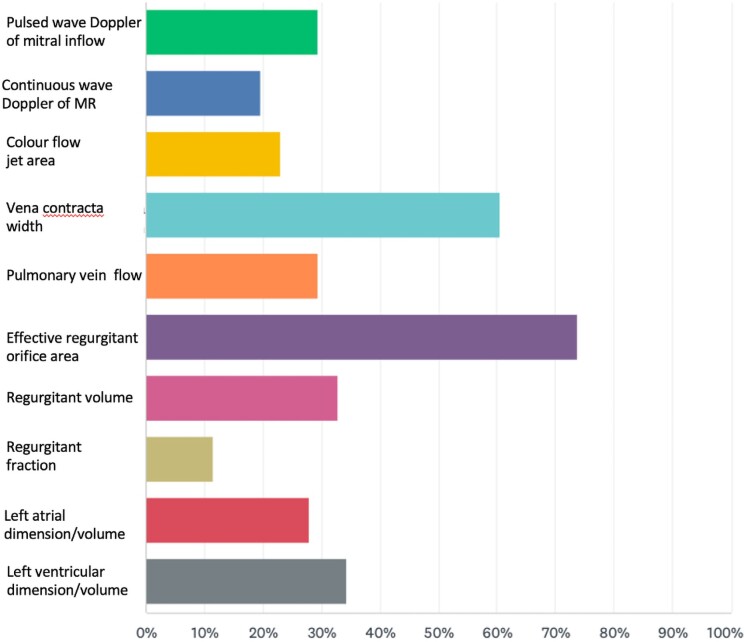
A total of 61 responders, mainly from tertiary centres or university hospitals, from 26 different countries responded to the survey, which consisted of 22 questions. For most questions related to echocardiography and advanced imaging, the answers were relatively homogeneous and demonstrated good adherence to current recommendations. In particular, the centres used a multi-parametric echocardiographic approach and selected the effective regurgitant orifice and vena contracta width as their preferred assessments. 2D measurements are still the most widely used parameters to assess left ventricular structure; however, the majority use 3D trans-oesophageal echocardiography (TOE) to evaluate valve morphology in severe MR. The majority of centres reported the onsite availability and clinical use of ergometric stress echocardiography, cardiac computed tomography (CCT), and cardiac magnetic resonance (CMR) imaging. Heart valve clinics and heart valve teams were also widely prevalent.
Consistent with current guidelines, echocardiography (transthoracic echocardiography and TOE) remains the first-line and central imaging modality for the assessment of MR although the complementary use of 3D TOE, CCT, and CMR appears to be growing. Heart valve clinics and heart valve teams are now widely prevalent.
评估欧洲常规临床实践中二尖瓣反流(MR)患者的诊断和影像学表现,以及管理方法。为此,欧洲心血管成像科学倡议委员会对欧洲各中心进行了调查。特别是,调查了超声心动图、高级影像学检查、心脏瓣膜门诊和心脏瓣膜团队的常规应用。
共有来自 26 个不同国家的 61 名应答者(主要来自三级中心或大学医院)回答了该调查,调查共包括 22 个问题。对于大多数与超声心动图和高级影像学相关的问题,答案相对一致,且显示出对当前建议的良好依从性。特别是,各中心采用了多参数超声心动图方法,并选择有效反流口和收缩期瓣口宽度作为首选评估指标。二维测量仍然是评估左心室结构最广泛使用的参数,但大多数中心使用 3 维经食管超声心动图(TOE)来评估严重 MR 中的瓣膜形态。大多数中心报告了现场可用性和临床应用运动负荷超声心动图、心脏计算机断层扫描(CCT)和心脏磁共振(CMR)成像。心脏瓣膜门诊和心脏瓣膜团队也广泛存在。
与当前指南一致,超声心动图(经胸超声心动图和 TOE)仍然是评估 MR 的一线和核心影像学方法,尽管 3 维 TOE、CCT 和 CMR 的补充应用似乎正在增加。心脏瓣膜门诊和心脏瓣膜团队现在已经广泛存在。