Department of Nuclear Medicine, Saarland University- Medical Center, Kirrberger Str. 100, Geb. 50, D-66421, Homburg, Germany.
Spencer-Fontayne Corporation, Jersey City, NJ, USA.
Cancer Imaging. 2024 Feb 22;24(1):27. doi: 10.1186/s40644-024-00671-1.
The state-of-the-art method for imaging men with biochemical recurrence of prostate cancer (BCR) is prostate-specific membrane antigen (PSMA)-targeted positron emission tomography/computed tomography (PET/CT) with tracers containing short-lived radionuclides, e.g., gallium-68 (Ga; half-life: ∼67.7 min). However, such imaging not infrequently yields indeterminate findings, which remain challenging to characterize. PSMA-targeted tracers labeled with zirconium-89 (Zr; half-life: ∼78.41 h) permit later scanning, which may help in classifying the level of suspiciousness for prostate cancer of lesions previously indeterminate on conventional PSMA-targeted PET/CT.
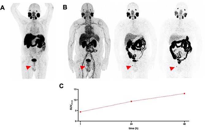
To assess the ability of [Zr]Zr-PSMA-617 PET/CT to characterize such lesions, we retrospectively analyzed altogether 20 lesions that were indeterminate on prior [Ga]Ga-PSMA-11 PET/CT, in 15 men with BCR (median prostate-specific antigen: 0.70 ng/mL). The primary endpoint was the lesions' classifications, and secondary endpoints included [Zr]Zr-PSMA-617 uptake (maximum standardized uptake value [SUV]), and lesion-to-background ratio (tumor-to-liver ratio of the SUV [TLR]). [Zr]Zr-PSMA-617 scans were performed 1 h, 24 h, and 48 h post-injection of 123 ± 19 MBq of radiotracer, 35 ± 35 d post-[Ga]Ga-PSMA-11 PET/CT.
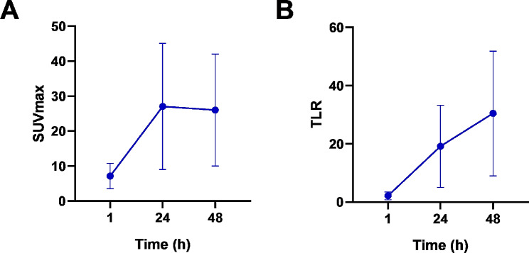
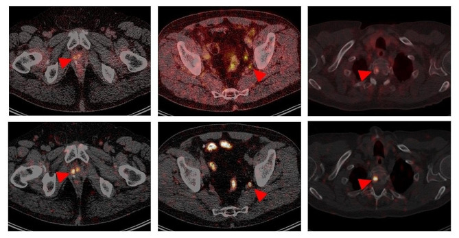
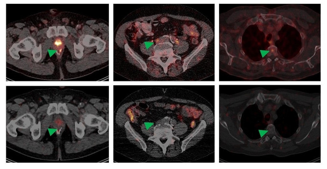
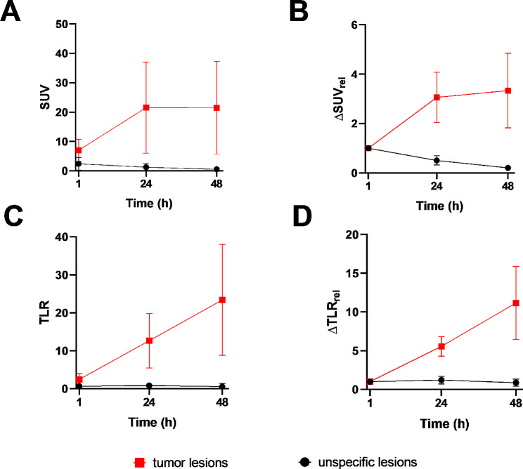
Altogether, 6/20 previously-indeterminate lesions (30%) were classified as suspicious (positive) for prostate cancer, 14/20 (70%), as non-suspicious (negative). In these two categories, [Zr]Zr-PSMA-617 uptake and lesional contrast showed distinctly different patterns. In positive lesions, SUV and TLR markedly rose from 1 to 48 h, with SUV essentially plateauing at high levels, and TLR further steeply increasing, from 24 to 48 h. In negative lesions, uptake, when present, was very low, and decreasing, while contrast was minimal, from 1 to 48 h. No adverse events or clinically-relevant vital signs changes related to [Zr]Zr-PSMA-617 PET/CT were noted during or ~ 4 weeks after the procedure.
In men with BCR, [Zr]Zr-PSMA-617 PET/CT may help characterize as suspicious or non-suspicious for prostate cancer lesions that were previously indeterminate on [Ga]Ga-PSMA-11 PET/CT.
Not applicable.
目前,用于成像生化复发前列腺癌(BCR)男性的最先进方法是使用含有短半衰期放射性核素的前列腺特异性膜抗原(PSMA)靶向正电子发射断层扫描/计算机断层扫描(PET/CT),例如镓-68(Ga;半衰期:67.7 min)。然而,这种成像并不罕见地会产生不确定的结果,这些结果仍然难以定性。用锆-89(Zr;半衰期:78.41 h)标记的 PSMA 靶向示踪剂允许进行延迟扫描,这可能有助于对之前在常规 PSMA 靶向 PET/CT 上不确定的病变进行可疑前列腺癌程度的分类。
为了评估 [Zr]Zr-PSMA-617 PET/CT 对这些病变的定性能力,我们回顾性分析了 15 名 BCR 男性中 20 个先前在 [Ga]Ga-PSMA-11 PET/CT 上不确定的病变(中位前列腺特异性抗原:0.70 ng/mL)。主要终点是病变的分类,次要终点包括 [Zr]Zr-PSMA-617 摄取(最大标准化摄取值 [SUV])和病变与背景比(SUV 的肿瘤与肝脏比 [TLR])。在 [Ga]Ga-PSMA-11 PET/CT 后 35 ± 35 d,注射 123 ± 19 MBq 放射性示踪剂后 1、24 和 48 h 进行 [Zr]Zr-PSMA-617 扫描。
总共,20 个先前不确定的病变中有 6/20(30%)被归类为可疑(阳性)前列腺癌,14/20(70%)为非可疑(阴性)。在这两个类别中,[Zr]Zr-PSMA-617 摄取和病变对比度显示出明显不同的模式。在阳性病变中,SUV 和 TLR 从 1 小时到 48 小时显著升高,SUV 基本保持在高水平,TLR 进一步急剧升高,从 24 小时到 48 小时。在阴性病变中,摄取(如果存在)非常低且逐渐减少,而对比度则从 1 小时到 48 小时非常低。在程序期间或程序后约 4 周内,未观察到与 [Zr]Zr-PSMA-617 PET/CT 相关的不良事件或临床相关生命体征变化。
在 BCR 男性中,[Zr]Zr-PSMA-617 PET/CT 可能有助于对 [Ga]Ga-PSMA-11 PET/CT 上先前不确定的病变进行可疑或非可疑的前列腺癌分类。
不适用。