Bhargava Mona, Bhambhani Varsha, Sen Ahana, Johri Aditi
Department of Cornea, Aditya Birla Sankara Nethralaya, 147/1 E M Bypass Road, Kolkata, 700099, India.
Aditya Birla Sankara Nethralaya, 147/1 E M Bypass Road, Kolkata, 700099, India.
Am J Ophthalmol Case Rep. 2024 Mar 14;34:102038. doi: 10.1016/j.ajoc.2024.102038. eCollection 2024 Jun.
To report a case of metallic corneal foreign-body (CFB) penetrating the Laser in situ keratomileusis (LASIK) flap and its successful outcome. To highlight usefulness of Anterior Segment Optical Coherence Tomography (ASOCT) in diagnosis and management of post-LASIK CFB. To enumerate other similar cases published in literature.
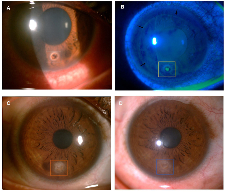
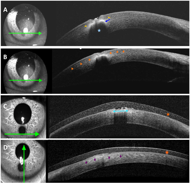
A 30-year-old male presented to the emergency department of a tertiary eye care centre with a metallic CFB. He had undergone uneventful LASIK elsewhere 4-years back. He was unaware of any trauma. CFB removal was attempted elsewhere but abandoned as CFB appeared deeply embedded. ASOCT showed CFB had penetrated LASIK flap and lodged into midstroma, 207 μm deep. CFB was successfully removed in operation theatre along with the application of cyanoacrylate glue and bandage contact lens. A review of literature for CFB in post-LASIK patients was done through PubMed search.
Postoperative course was uncomplicated and there was a follow up period of 4 months. Vision improved to unaided 20/20 and N/6 from preoperative 20/60 and N/10. Review of literature of 24 patients showed Post-LASIK FB was more common in males (79%). None of the patients except for one had protective eye-wear. Metallic FB was most common followed by organic FB. Flap complications were present in seven patients. Diffuse lamellar keratitis (DLK) and epithelial ingrowth were the most common post-FB removal complications occurring in six (25%) and four (16.6%) patients respectively.
Post-LASIK patients with CFB need to be inspected for flap related complications. CFB can be successfully removed, although DLK, epithelial ingrowth, microbial keratitis, astigmatism, can occur post-CFB removal. ASOCT can delineate CFB and flap related details and thus is an additional useful imaging tool in such scenarios.
报告一例金属性角膜异物(CFB)穿透准分子原位角膜磨镶术(LASIK)瓣并取得成功治疗结果的病例。强调眼前节光学相干断层扫描(ASOCT)在LASIK术后CFB诊断和处理中的作用。列举文献中报道的其他类似病例。
一名30岁男性因金属性CFB就诊于一家三级眼科护理中心的急诊科。他4年前在其他地方接受了顺利的LASIK手术。他不知道有任何外伤史。在其他地方尝试取出CFB,但因CFB似乎深深嵌入而放弃。ASOCT显示CFB穿透了LASIK瓣并嵌入基质中层,深度为207μm。在手术室成功取出CFB,并应用了氰基丙烯酸酯胶水和绷带式隐形眼镜。通过PubMed搜索对LASIK术后患者CFB的文献进行了回顾。
术后过程无并发症,随访期为4个月。视力从术前的20/60和N/10提高到术后裸眼20/20和N/6。对24例患者的文献回顾显示,LASIK术后异物在男性中更常见(79%)。除1例患者外,其他患者均未佩戴防护眼镜。金属性异物最常见,其次是有机异物。7例患者出现瓣相关并发症。弥漫性板层角膜炎(DLK)和上皮植入是异物取出后最常见的并发症,分别发生在6例(25%)和4例(16.6%)患者中。
LASIK术后出现CFB的患者需要检查是否有瓣相关并发症。CFB可以成功取出,尽管在CFB取出后可能会发生DLK、上皮植入、微生物性角膜炎、散光。ASOCT可以描绘CFB和瓣相关细节,因此在这种情况下是一种额外有用的成像工具。