Han Huan, Yin Shu-Yi, Song Rui-Xiang, Zhao Jing, Yu Yong-Wei, He Miao-Xia, Wang Han
Department of Pathology, Changhai Hospital, Naval Medical University, Shanghai, China.
Department of Urology, Changhai Hospital, Naval Medical University, Shanghai, China.
Transl Androl Urol. 2024 Mar 31;13(3):383-396. doi: 10.21037/tau-23-518. Epub 2024 Mar 18.
Papillary renal neoplasm with reverse polarity (PRNRP) is a novel entity with unique clinicopathological characteristics, and only a small number of patients with PRNRP have been described.
We retrospectively analyzed the data for nine patients with PRNRP and evaluated differences in the clinical, histomorphological, immunohistochemical, and molecular features; prognosis; and differential diagnosis of PRNRP from other renal tumors with papillary structure.
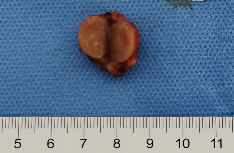
There were six males and three females aged 36 to 74 years (mean: 62.33 years; median: 68 years). All the tumors were solitary and ranged from 1 to 3.7 cm (mean: 2.17 cm; median: 2 cm), with three and six tumors arose in the left and right renal tract, respectively. Pathologically, PRNRP is a small, well-circumscribed neoplasm with predominant papillary formations. The lining epithelium is composed of a monolayer of cuboidal to low-columnar cells with low-grade nuclei arranged against the apical pole of the tumor cells. Edema, mucinous degeneration, and hyaline degeneration are found in the fibrovascular cores. Foamy macrophages, psammoma bodies, hemosiderin deposition, and infiltrative tumor boundaries were present in some patients. Immunohistochemically, all tumors showed diffuse positive staining for GATA3. Sanger sequencing confirmed the presence of mutation in seven patients. All patients had a good prognosis after surgery and were relapse free. Positive staining for GATA3 and negative staining for vimentin were the most significant markers for differentiating PRNRP from other renal tumors with analogous structure.
These findings suggested that PRNRP is a distinctive subtype of renal tumor with specific pathological features and indolent behaviors that should be distinguished from other renal tumors, especially papillary renal cell carcinoma. A monolayer of tumor cells with an inverted nuclear pattern, positive staining for , and KRAS mutation are essential for pathological diagnosis. Owing to its satisfactory prognosis, the surveillance and follow-up of patients with PRNRP should be additionally formulated.
具有反向极性的乳头状肾肿瘤(PRNRP)是一种具有独特临床病理特征的新型实体,仅有少数PRNRP患者被报道。
我们回顾性分析了9例PRNRP患者的数据,并评估了其临床、组织形态学、免疫组化和分子特征、预后以及PRNRP与其他具有乳头状结构的肾肿瘤的鉴别诊断。
9例患者中男性6例,女性3例,年龄36至74岁(平均62.33岁;中位数68岁)。所有肿瘤均为单发,大小为1至3.7 cm(平均2.17 cm;中位数2 cm),其中左肾和右肾分别有3例和6例肿瘤。病理上,PRNRP是一种边界清楚的小肿瘤,主要呈乳头状结构。衬里上皮由单层立方至低柱状细胞组成,细胞核级别低,排列于肿瘤细胞的顶端极。纤维血管核心可见水肿、黏液样变性和玻璃样变性。部分患者可见泡沫状巨噬细胞、砂粒体、含铁血黄素沉积及浸润性肿瘤边界。免疫组化显示,所有肿瘤GATA3均呈弥漫性阳性染色。桑格测序证实7例患者存在突变。所有患者术后预后良好,无复发。GATA3阳性染色和波形蛋白阴性染色是PRNRP与其他具有类似结构的肾肿瘤鉴别的最重要标志物。
这些发现提示,PRNRP是一种具有独特病理特征和惰性生物学行为的肾肿瘤独特亚型,应与其他肾肿瘤尤其是乳头状肾细胞癌相鉴别。具有核倒转模式的单层肿瘤细胞、GATA3阳性染色及KRAS突变对病理诊断至关重要。鉴于其预后良好,应额外制定PRNRP患者的监测和随访方案。