1st Orthopaedic and Traumatologic Clinic, IRCCS Istituto Ortopedico Rizzoli, Via G.B. Pupilli 1, Bologna, 40136, Italy.
Arch Orthop Trauma Surg. 2024 May;144(5):2305-2316. doi: 10.1007/s00402-024-05332-3. Epub 2024 Apr 20.
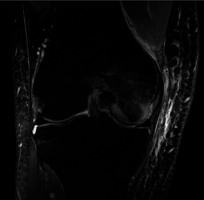
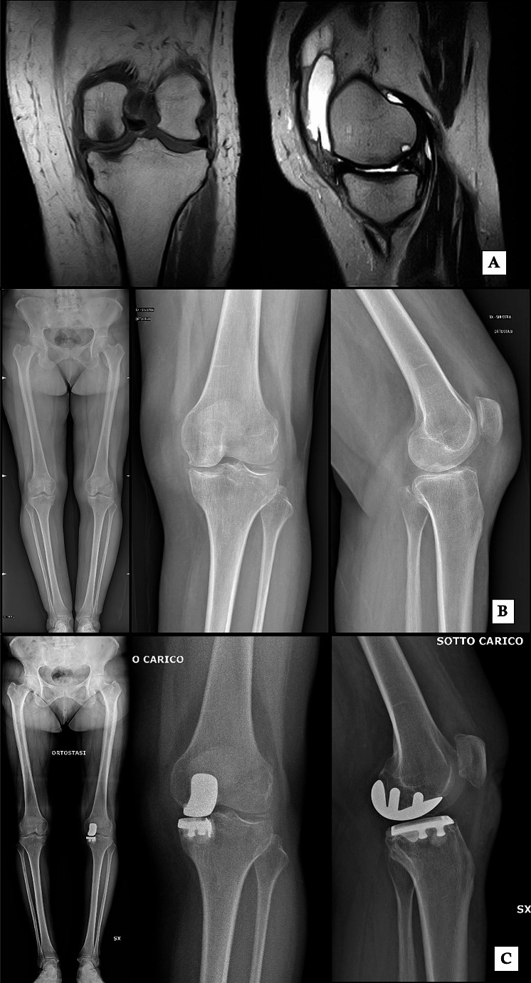
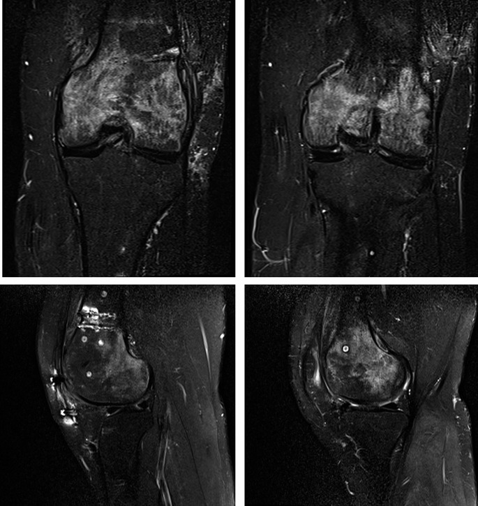
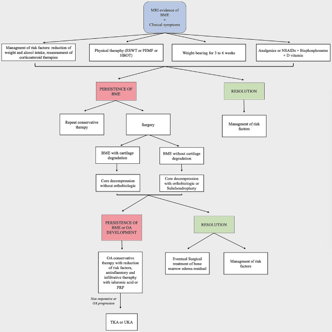
Bone marrow edema (BME) is a frequent MRI finding in patients with knee pain. According to the etiology, BME of the knee can be classified into three main categories: ischemic, mechanic, and reactive. The diagnosis may be difficult, because of the specificity of symptoms and the poor radiographic findings. MRI is the gold standard, showing an area of altered signal of the bone with an high signal intensity on fat-suppressed, T2 weighted images, usually in combination with an intermediate or low signal intensity on T1 weighted images. Bone marrow edema tends to be self-limiting and, in most cases, resolves without any consequences in a varying amount of time. However, since it may evolve to complete joint destruction, early diagnosis and correct treatment are crucial to prevent the articular degeneration. Conservative therapy is the first step, with no weight-bearing for 3 to 6 weeks on the affected side, in combination with the administration of anti-inflammatory drugs or painkillers to manage symptoms. In non-responding forms and more advanced stages, minimally invasive preservative surgery can provide significant results, with subchondroplasty and core decompression being the two main procedures available. Knee arthroplasty, both total (TKA) or unicompartmental (UKA), is the only effective option when the degradation of cartilage is diffuse and in patients with subchondral bone collapse.
骨髓水肿(BME)是膝关节疼痛患者常见的 MRI 表现。根据病因,膝关节的 BME 可分为三大类:缺血性、机械性和反应性。由于症状的特异性和影像学表现不佳,诊断可能具有挑战性。MRI 是金标准,显示骨信号改变区,在脂肪抑制 T2 加权图像上呈高信号强度,通常在 T1 加权图像上呈中等或低信号强度。骨髓水肿往往具有自限性,在大多数情况下,在不同时间内无需任何后果即可自行消退。然而,由于它可能发展为完全关节破坏,因此早期诊断和正确治疗对于预防关节退变至关重要。保守治疗是第一步,患侧 3 至 6 周不负重,并结合使用抗炎药或止痛药来缓解症状。在非反应性形式和更晚期,微创保膝手术可以提供显著的效果,其中软骨下成形术和核心减压术是两种主要的手术方法。当软骨降解弥漫且存在软骨下骨塌陷时,膝关节置换术(TKA 或 UKA)是唯一有效的选择。