• 文献检索
  • 文档翻译
  • 深度研究
  • 学术资讯
  • Suppr Zotero 插件Zotero 插件
  • 邀请有礼
  • 套餐&价格
  • 历史记录
应用&插件
Suppr Zotero 插件Zotero 插件浏览器插件Mac 客户端Windows 客户端微信小程序
定价
高级版会员购买积分包购买API积分包
服务
文献检索文档翻译深度研究API 文档MCP 服务
关于我们
关于 Suppr公司介绍联系我们用户协议隐私条款
关注我们

Suppr 超能文献

核心技术专利:CN118964589B侵权必究
粤ICP备2023148730 号-1Suppr @ 2026

文献检索

告别复杂PubMed语法,用中文像聊天一样搜索,搜遍4000万医学文献。AI智能推荐,让科研检索更轻松。

立即免费搜索

文件翻译

保留排版,准确专业,支持PDF/Word/PPT等文件格式,支持 12+语言互译。

免费翻译文档

深度研究

AI帮你快速写综述,25分钟生成高质量综述,智能提取关键信息,辅助科研写作。

立即免费体验

梅尼埃病患者计算机断层扫描与磁共振成像的前庭导水管可视化比较

Comparison of vestibular aqueduct visualization on computed tomography and magnetic resonance imaging in patients with Ménière's disease.

作者信息

Xia Kaijun, Lei Ping, Liu Yingzhao, Chen Cen, Pan Hui, Leng Yangming, Liu Bo

机构信息

Department of Otorhinolaryngology-Head and Neck Surgery, Union Hospital, Tongji Medical College, Huazhong University of Science and Technology, Wuhan, 430022, China.

Department of Radiology, Union Hospital, Tongji Medical College, Huazhong University of Science and Technology, Wuhan, 430022, China.

出版信息

BMC Med Imaging. 2024 Apr 22;24(1):93. doi: 10.1186/s12880-024-01275-8.

DOI:10.1186/s12880-024-01275-8
PMID:38649991
原文链接:https://pmc.ncbi.nlm.nih.gov/articles/PMC11034041/
Abstract

BACKGROUND

The vestibular aqueduct (VA) serves an essential role in homeostasis of the inner ear and pathogenesis of Ménière's disease (MD). The bony VA can be clearly depicted by high-resolution computed tomography (HRCT), whereas the optimal sequences and parameters for magnetic resonance imaging (MRI) are not yet established. We investigated VA characteristics and potential factors influencing MRI-VA visibility in unilateral MD patients.

METHODS

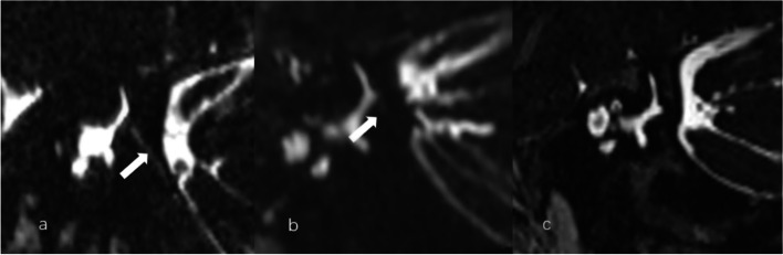
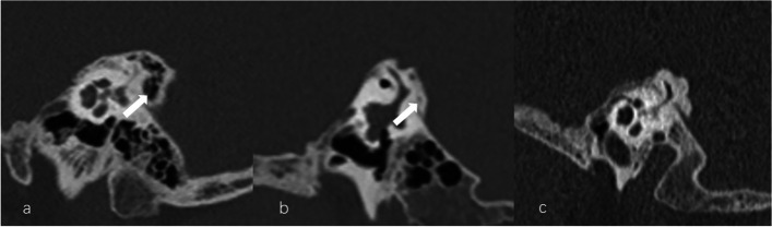
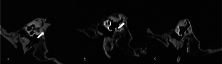
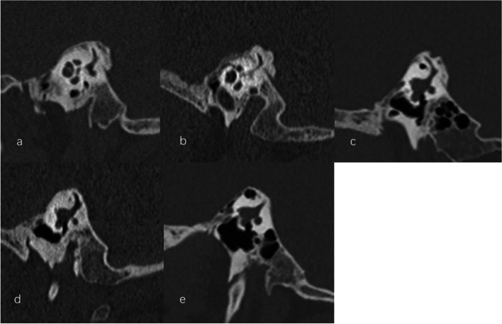
One hundred patients with unilateral MD underwent MRI with three-dimensional sampling perfection with application optimized contrasts using different flip angle evolutions (3D-SPACE) sequence and HRCT evaluation. The imaging variables included MRI-VA and CT-VA visibility, CT-VA morphology and CT-peri-VA pneumatization.

RESULTS

The most frequent type of MRI-VA and CT-VA visualization was invisible VA and continuous VA, respectively. The MRI-VA visibility was significantly lower than CT-VA visibility. MRI-VA visibility had a weak positive correlation with ipsilateral CT-VA visualization. For the affected side, the MRI-VA visualization was negatively correlated with the incidence of obliterated-shaped CT-VA and positively with that of tubular-shaped CT-VA. MRI-VA visualization was not affected by CT-peri-VA pneumatization.

CONCLUSION

In patients with MD, the VA visualization on 3D-SPACE MRI is poorer than that observed on CT and may be affected by its osseous configuration. These findings may provide a basis for further characterization of VA demonstrated by MRI and its clinical significance.

摘要

背景

前庭导水管(VA)在内耳稳态及梅尼埃病(MD)发病机制中起重要作用。高分辨率计算机断层扫描(HRCT)能清晰显示骨性VA,而磁共振成像(MRI)的最佳序列和参数尚未确定。我们研究了单侧MD患者的VA特征及影响MRI-VA可见性的潜在因素。

方法

100例单侧MD患者接受了使用不同翻转角演化的三维采样完美应用优化对比(3D-SPACE)序列的MRI检查及HRCT评估。成像变量包括MRI-VA和CT-VA的可见性、CT-VA形态及CT-VA周围气化情况。

结果

MRI-VA和CT-VA最常见的可视化类型分别为不可见VA和连续VA。MRI-VA的可见性显著低于CT-VA的可见性。MRI-VA的可见性与同侧CT-VA的可视化呈弱正相关。对于患侧,MRI-VA的可视化与闭塞型CT-VA的发生率呈负相关,与管状CT-VA的发生率呈正相关。MRI-VA的可视化不受CT-VA周围气化的影响。

结论

在MD患者中,3D-SPACE MRI上VA的可视化比CT上观察到的要差,且可能受其骨质结构影响。这些发现可能为进一步表征MRI显示的VA及其临床意义提供依据。

https://cdn.ncbi.nlm.nih.gov/pmc/blobs/5af2/11034041/291c392d28a2/12880_2024_1275_Fig4_HTML.jpg
https://cdn.ncbi.nlm.nih.gov/pmc/blobs/5af2/11034041/33e79da589a8/12880_2024_1275_Fig1_HTML.jpg
https://cdn.ncbi.nlm.nih.gov/pmc/blobs/5af2/11034041/46d67993c252/12880_2024_1275_Fig2_HTML.jpg
https://cdn.ncbi.nlm.nih.gov/pmc/blobs/5af2/11034041/aefb3ca56cf1/12880_2024_1275_Fig3_HTML.jpg
https://cdn.ncbi.nlm.nih.gov/pmc/blobs/5af2/11034041/291c392d28a2/12880_2024_1275_Fig4_HTML.jpg

相似文献

1
Comparison of vestibular aqueduct visualization on computed tomography and magnetic resonance imaging in patients with Ménière's disease.梅尼埃病患者计算机断层扫描与磁共振成像的前庭导水管可视化比较
BMC Med Imaging. 2024 Apr 22;24(1):93. doi: 10.1186/s12880-024-01275-8.
2
The vestibular aqueduct ossification on temporal bone CT: an old sign revisited to rule out the presence of endolymphatic hydrops in Menière's disease patients.颞骨 CT 显示前庭水管骨化:重新审视这一古老征象,以排除梅尼埃病患者存在内淋巴积水。
Eur Radiol. 2020 Nov;30(11):6331-6338. doi: 10.1007/s00330-020-06980-w. Epub 2020 Jun 15.
3
Value of CT and three-dimensional reconstruction revealing specific radiological signs for screening causative high jugular bulb in patients with Meniere's disease.CT 及三维重建对梅尼埃病患者高位颈静脉球显示特定影像学特征的筛查价值。
BMC Med Imaging. 2020 Aug 31;20(1):103. doi: 10.1186/s12880-020-00504-0.
4
Relationship between multidetector CT imaging of the vestibular aqueduct and inner ear pathologies.前庭导水管的多排螺旋CT成像与内耳病变之间的关系。
Neuroradiol J. 2013 Dec;26(6):683-92. doi: 10.1177/197140091302600612. Epub 2013 Dec 18.
5
The vestibular aqueduct sign: Magnetic resonance imaging can detect abnormalities in both ears of patients with unilateral Meniere's disease.前庭水管征:磁共振成像可检测单侧梅尼埃病患者双耳的异常。
J Neuroradiol. 2020 Mar;47(2):174-179. doi: 10.1016/j.neurad.2018.10.003. Epub 2018 Nov 10.
6
Practical 3DCT imaging of the vestibular aqueduct for Meniere's disease.梅尼埃病的前庭导水管实用三维CT成像
Acta Otolaryngol. 2015 Aug;135(8):799-806. doi: 10.3109/00016489.2015.1034879. Epub 2015 Apr 25.
7
Assessing the optimal MRI descriptors to diagnose Ménière's disease and the added value of analysing the vestibular aqueduct.评估诊断梅尼埃病的最佳 MRI 描述符及分析前庭水管的附加价值。
Eur Radiol. 2024 Sep;34(9):6060-6071. doi: 10.1007/s00330-024-10587-w. Epub 2024 Feb 7.
8
Anatomical variation of inner ear may be a predisposing factor for unilateral Ménière's disease rather than for ipsilateral delayed endolymphatic hydrops.内耳解剖结构的变异可能是单侧梅尼埃病的一个易感因素,而不是同侧迟发性内淋巴积水的易感因素。
Eur Radiol. 2022 May;32(5):3553-3564. doi: 10.1007/s00330-021-08430-7. Epub 2022 Jan 3.
9
Association Between Vestibular Aqueduct Morphology and Meniere's Disease.前庭水管形态与梅尼埃病的关系。
Laryngoscope. 2024 Jul;134(7):3349-3354. doi: 10.1002/lary.31339. Epub 2024 Feb 17.
10
Non-contrast MRI of Inner Ear Detected Differences of Endolymphatic Drainage System Between Vestibular Migraine and Unilateral Ménière's Disease.内耳非增强磁共振成像检测前庭性偏头痛与单侧梅尼埃病内淋巴引流系统的差异
Front Neurol. 2022 Apr 29;13:814518. doi: 10.3389/fneur.2022.814518. eCollection 2022.

引用本文的文献

1
Angular Trajectory of the Vestibular Aqueduct in a Cohort of Chinese Patients with Unilateral Ménière's Disease: Association with Other Imaging Indices and Clinical Profiles.一组中国单侧梅尼埃病患者前庭导水管的角轨迹:与其他影像学指标及临床特征的关联
Biomedicines. 2024 Sep 3;12(9):2008. doi: 10.3390/biomedicines12092008.

本文引用的文献

1
Does CISS MRI Reliably Depict the Endolymphatic Duct in Children with and without Vestibular Aqueduct Enlargement?CISS MRI 是否能可靠地显示伴有和不伴有前庭导水管扩大的儿童内淋巴管?
AJNR Am J Neuroradiol. 2024 Apr 8;45(4):511-517. doi: 10.3174/ajnr.A8158.
2
Assessing the optimal MRI descriptors to diagnose Ménière's disease and the added value of analysing the vestibular aqueduct.评估诊断梅尼埃病的最佳 MRI 描述符及分析前庭水管的附加价值。
Eur Radiol. 2024 Sep;34(9):6060-6071. doi: 10.1007/s00330-024-10587-w. Epub 2024 Feb 7.
3
Visualization and clinical relevance of the endolymphatic duct and sac in Ménière's disease.
梅尼埃病中内淋巴管和内淋巴囊的可视化及其临床相关性
Front Neurol. 2023 Aug 31;14:1239422. doi: 10.3389/fneur.2023.1239422. eCollection 2023.
4
Comparison of radiological abnormalities between the jugular bulb and the vestibular aqueduct in patients with Ménière's disease.梅尼埃病患者颈静脉球与前庭导水管之间放射学异常的比较。
Front Neurol. 2023 May 12;14:1184232. doi: 10.3389/fneur.2023.1184232. eCollection 2023.
5
Meniere's Disease Patients with Distinct Sac Pathoanatomic Findings React Differently to Endolymphatic Duct Blockage.梅尼埃病患者的囊腔病理改变不同,对内淋巴囊阻塞的反应也不同。
Laryngoscope. 2023 Oct;133(10):2761-2769. doi: 10.1002/lary.30520. Epub 2022 Dec 19.
6
Non-contrast MRI of Inner Ear Detected Differences of Endolymphatic Drainage System Between Vestibular Migraine and Unilateral Ménière's Disease.内耳非增强磁共振成像检测前庭性偏头痛与单侧梅尼埃病内淋巴引流系统的差异
Front Neurol. 2022 Apr 29;13:814518. doi: 10.3389/fneur.2022.814518. eCollection 2022.
7
Anatomical variation of inner ear may be a predisposing factor for unilateral Ménière's disease rather than for ipsilateral delayed endolymphatic hydrops.内耳解剖结构的变异可能是单侧梅尼埃病的一个易感因素,而不是同侧迟发性内淋巴积水的易感因素。
Eur Radiol. 2022 May;32(5):3553-3564. doi: 10.1007/s00330-021-08430-7. Epub 2022 Jan 3.
8
Vestibular Aqueduct Size Correlates With the Degree of Cochlear Hydrops in Patients With and Without Menière's Disease.前庭水管大小与伴有和不伴有梅尼埃病的患者耳蜗积水程度相关。
Otol Neurotol. 2021 Dec 1;42(10):e1532-e1536. doi: 10.1097/MAO.0000000000003300.
9
Radiological Configuration of the Vestibular Aqueduct Predicts Bilateral Progression in Meniere's Disease.前庭导水管的放射学形态可预测梅尼埃病的双侧进展。
Front Neurol. 2021 Jun 8;12:674170. doi: 10.3389/fneur.2021.674170. eCollection 2021.
10
The vestibular aqueduct ossification on temporal bone CT: an old sign revisited to rule out the presence of endolymphatic hydrops in Menière's disease patients.颞骨 CT 显示前庭水管骨化:重新审视这一古老征象,以排除梅尼埃病患者存在内淋巴积水。
Eur Radiol. 2020 Nov;30(11):6331-6338. doi: 10.1007/s00330-020-06980-w. Epub 2020 Jun 15.