Ranji Parmida, Jonasson Emma, Andersson Lisa, Filges Stefan, Luna Santamaría Manuel, Vannas Christoffer, Dolatabadi Soheila, Gustafsson Anna, Myklebost Ola, Håkansson Joakim, Fagman Henrik, Landberg Göran, Åman Pierre, Ståhlberg Anders
Sahlgrenska Center for Cancer Research, Department of Laboratory Medicine, Institute of Biomedicine, Sahlgrenska Academy at University of Gothenburg, Gothenburg, Sweden.
Wallenberg Centre for Molecular and Translational Medicine, University of Gothenburg, Gothenburg, Sweden.
J Transl Med. 2024 Apr 26;22(1):389. doi: 10.1186/s12967-024-05211-w.
Myxoid liposarcoma (MLS) displays a distinctive tumor microenvironment and is characterized by the FUS::DDIT3 fusion oncogene, however, the precise functional contributions of these two elements remain enigmatic in tumor development.
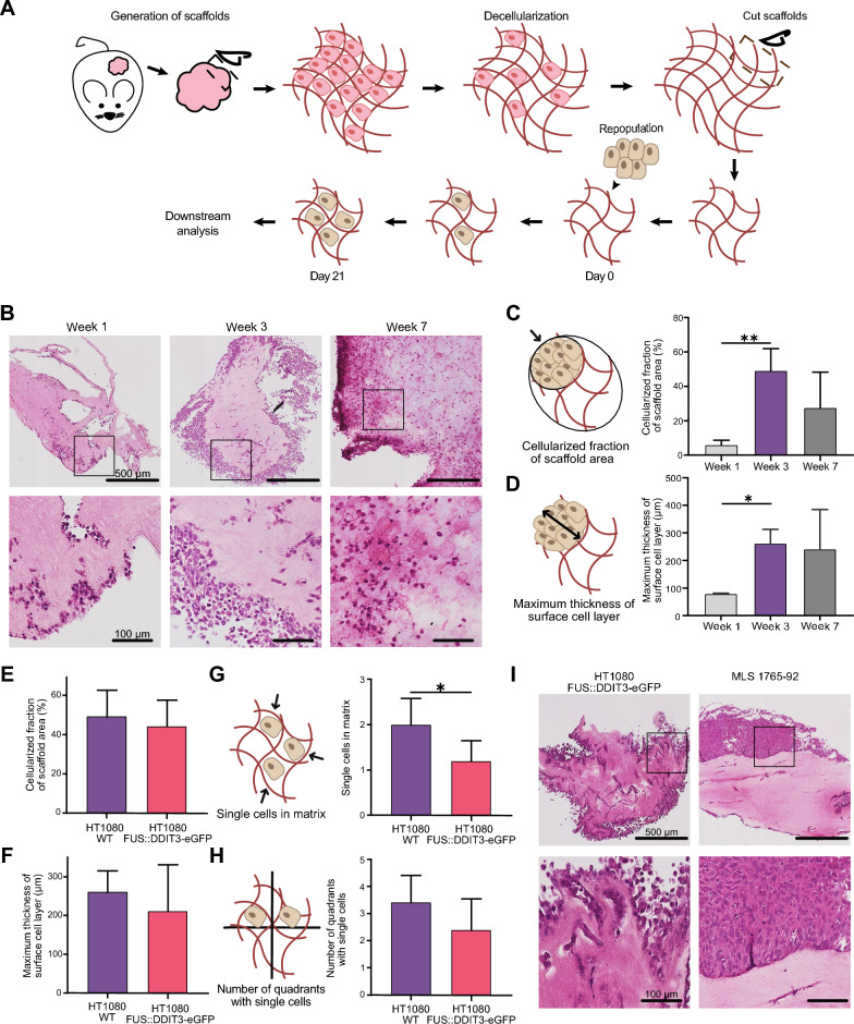
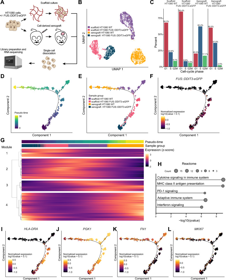
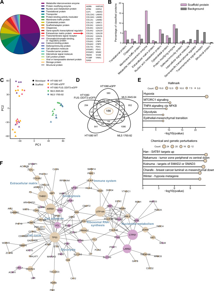
To study the cell-free microenvironment in MLS, we developed an experimental model system based on decellularized patient-derived xenograft tumors. We characterized the cell-free scaffold using mass spectrometry. Subsequently, scaffolds were repopulated using sarcoma cells with or without FUS::DDIT3 expression that were analyzed with histology and RNA sequencing.
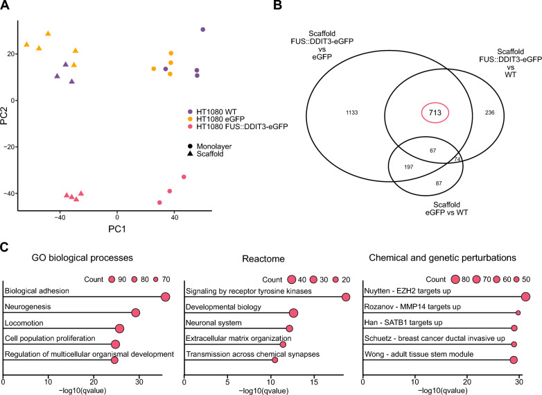
Characterization of cell-free MLS scaffolds revealed intact structure and a large variation of protein types remaining after decellularization. We demonstrated an optimal culture time of 3 weeks and showed that FUS::DDIT3 expression decreased cell proliferation and scaffold invasiveness. The cell-free MLS microenvironment and FUS::DDIT3 expression both induced biological processes related to cell-to-cell and cell-to-extracellular matrix interactions, as well as chromatin remodeling, immune response, and metabolism. Data indicated that FUS::DDIT3 expression more than the microenvironment determined the pre-adipocytic phenotype that is typical for MLS.
Our experimental approach opens new means to study the tumor microenvironment in detail and our findings suggest that FUS::DDIT3-expressing tumor cells can create their own extracellular niche.
黏液样脂肪肉瘤(MLS)具有独特的肿瘤微环境,其特征为FUS::DDIT3融合致癌基因,然而,这两个因素在肿瘤发展中的确切功能贡献仍不明确。
为研究MLS中的无细胞微环境,我们基于去细胞化的患者来源异种移植肿瘤开发了一个实验模型系统。我们使用质谱对无细胞支架进行了表征。随后,使用表达或不表达FUS::DDIT3的肉瘤细胞重新接种支架,并通过组织学和RNA测序进行分析。
对无细胞MLS支架的表征显示其结构完整,去细胞化后残留的蛋白质类型差异很大。我们证明了最佳培养时间为3周,并表明FUS::DDIT3表达降低了细胞增殖和支架侵袭性。无细胞MLS微环境和FUS::DDIT3表达均诱导了与细胞间和细胞与细胞外基质相互作用以及染色质重塑、免疫反应和代谢相关的生物学过程。数据表明,FUS::DDIT3表达比微环境更能决定MLS典型的前脂肪细胞表型。
我们的实验方法为详细研究肿瘤微环境开辟了新途径,我们的研究结果表明,表达FUS::DDIT3的肿瘤细胞可以创造自己的细胞外生态位。