文献检索文档翻译深度研究
Suppr Zotero 插件Zotero 插件
邀请有礼套餐&价格历史记录

新学期,新优惠

限时优惠:9月1日-9月22日

30天高级会员仅需29元

1天体验卡首发特惠仅需5.99元

了解详情
不再提醒
插件&应用
Suppr Zotero 插件Zotero 插件浏览器插件Mac 客户端Windows 客户端微信小程序
高级版
套餐订阅购买积分包
AI 工具
文献检索文档翻译深度研究
关于我们
关于 Suppr公司介绍联系我们用户协议隐私条款
关注我们

Suppr 超能文献

核心技术专利:CN118964589B侵权必究
粤ICP备2023148730 号-1Suppr @ 2025

血管内皮衍生的 SPARCL1 通过促炎巨噬细胞活化加重病毒性肺炎。

Vascular endothelial-derived SPARCL1 exacerbates viral pneumonia through pro-inflammatory macrophage activation.

机构信息

Department of Biomedical Sciences, School of Veterinary Medicine, University of Pennsylvania, Philadelphia, PA, 19104, USA.

Institute for Regenerative Medicine, University of Pennsylvania, Philadelphia, PA, 19104, USA.

出版信息

Nat Commun. 2024 May 18;15(1):4235. doi: 10.1038/s41467-024-48589-3.


DOI:10.1038/s41467-024-48589-3
PMID:38762489
原文链接:https://pmc.ncbi.nlm.nih.gov/articles/PMC11102455/
Abstract

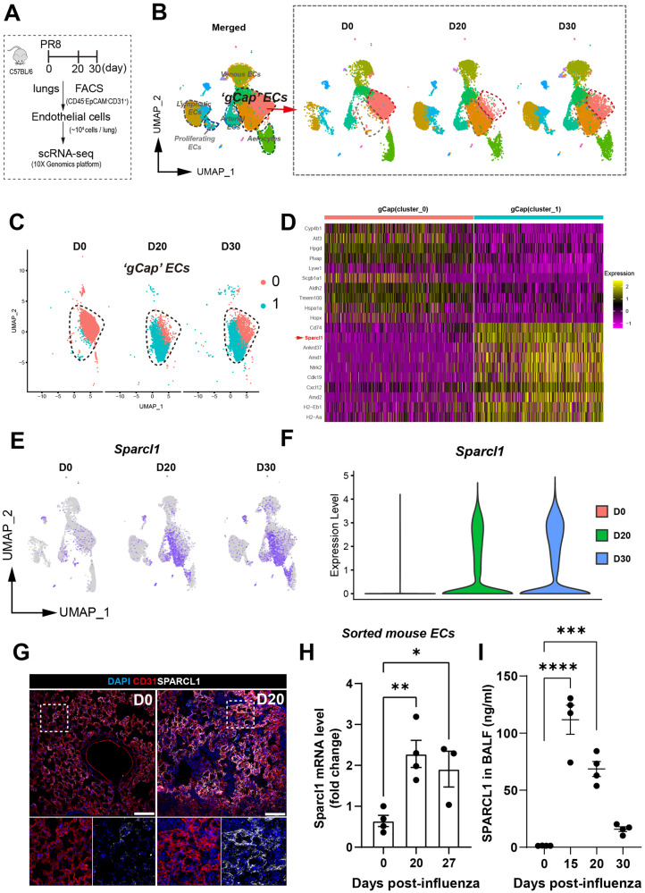
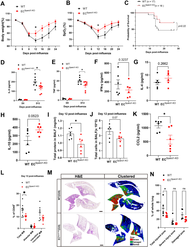
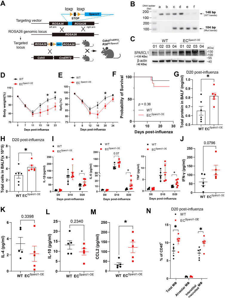
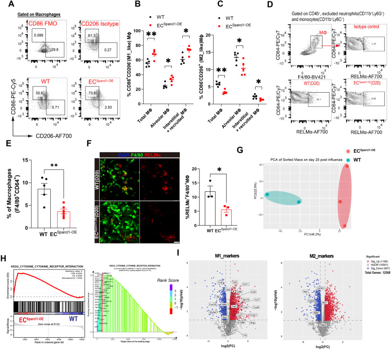
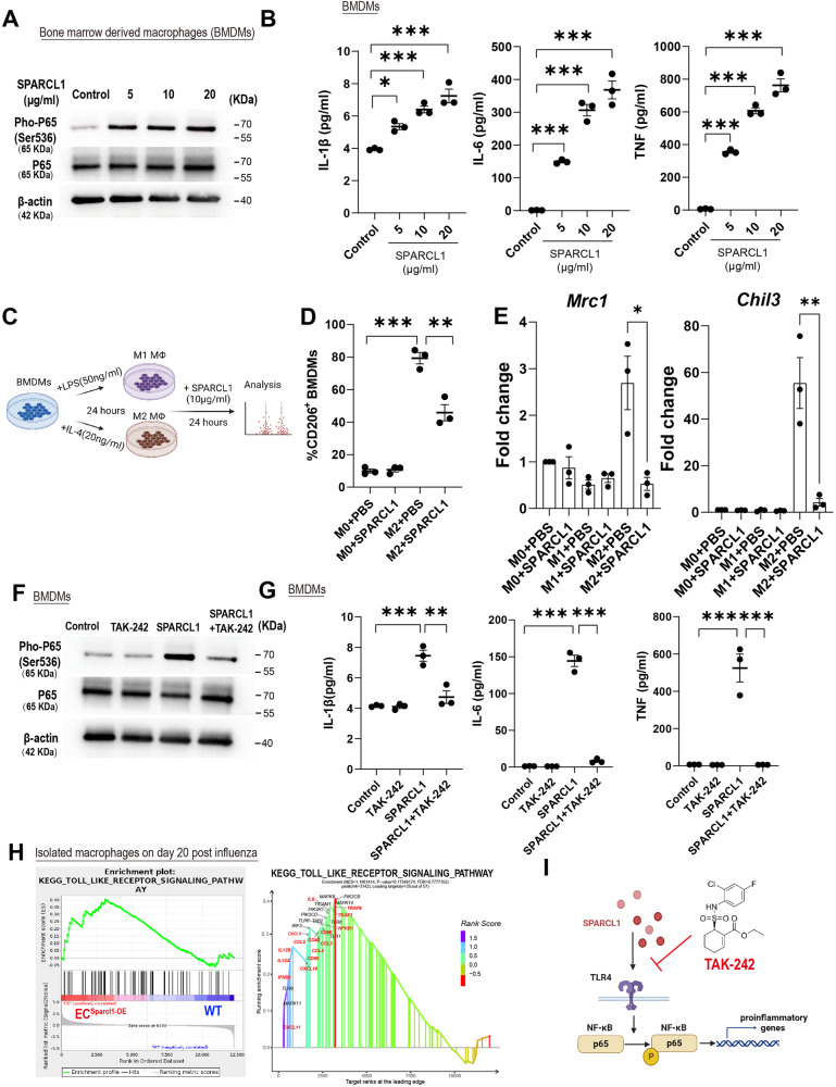
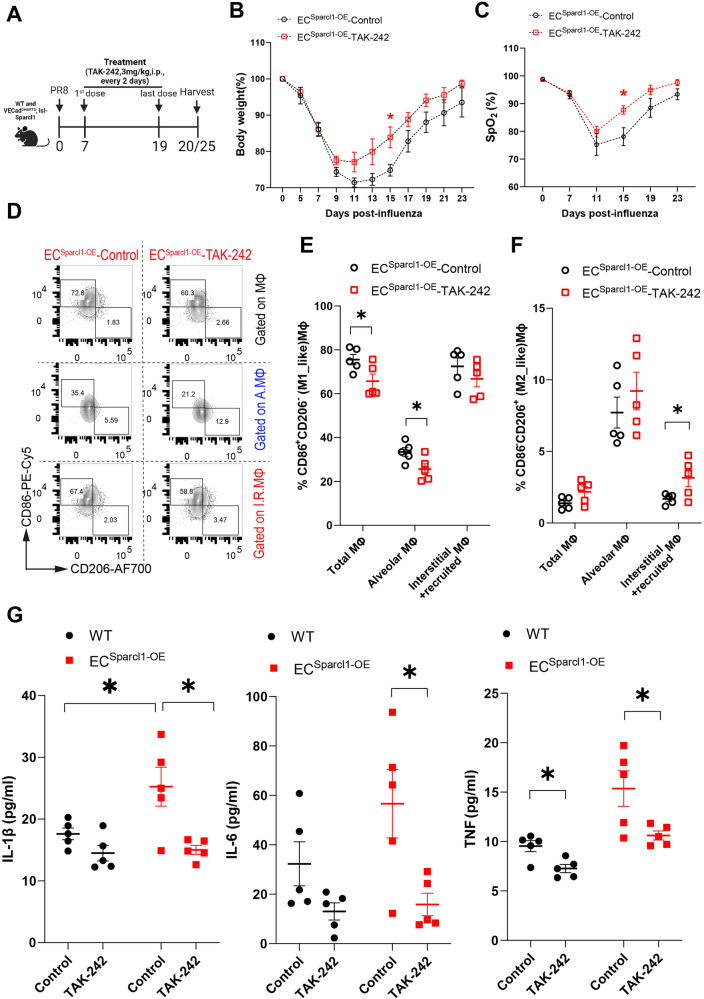
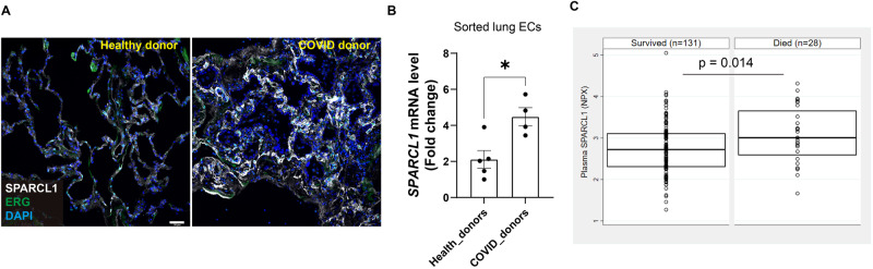
Inflammation induced by lung infection is a double-edged sword, moderating both anti-viral and immune pathogenesis effects; the mechanism of the latter is not fully understood. Previous studies suggest the vasculature is involved in tissue injury. Here, we report that expression of Sparcl1, a secreted matricellular protein, is upregulated in pulmonary capillary endothelial cells (EC) during influenza-induced lung injury. Endothelial overexpression of SPARCL1 promotes detrimental lung inflammation, with SPARCL1 inducing 'M1-like' macrophages and related pro-inflammatory cytokines, while SPARCL1 deletion alleviates these effects. Mechanistically, SPARCL1 functions through TLR4 on macrophages in vitro, while TLR4 inhibition in vivo ameliorates excessive inflammation caused by endothelial Sparcl1 overexpression. Finally, SPARCL1 expression is increased in lung ECs from COVID-19 patients when compared with healthy donors, while fatal COVID-19 correlates with higher circulating SPARCL1 protein levels in the plasma. Our results thus implicate SPARCL1 as a potential prognosis biomarker for deadly COVID-19 pneumonia and as a therapeutic target for taming hyperinflammation in pneumonia.

摘要

肺部感染引起的炎症是一把双刃剑,调节抗病毒和免疫发病机制的作用; 后者的机制尚未完全了解。先前的研究表明血管参与组织损伤。在这里,我们报告说,在流感诱导的肺损伤期间,分泌细胞外基质蛋白 Sparcl1 在肺毛细血管内皮细胞 (EC) 中上调。内皮细胞中 SPARCL1 的过表达促进有害的肺部炎症,SPARCL1 诱导“M1 样”巨噬细胞和相关的促炎细胞因子,而 SPARCL1 的缺失减轻了这些作用。从机制上讲,SPARCL1 在体外通过巨噬细胞上的 TLR4 发挥作用,而体内 TLR4 抑制可减轻内皮细胞 Sparcl1 过表达引起的过度炎症。最后,与健康供体相比,来自 COVID-19 患者的肺 EC 中 SPARCL1 的表达增加,而致命性 COVID-19 与血浆中循环 SPARCL1 蛋白水平升高相关。因此,我们的研究结果表明 SPARCL1 是致命性 COVID-19 肺炎的潜在预后生物标志物,并可作为控制肺炎过度炎症的治疗靶点。

https://cdn.ncbi.nlm.nih.gov/pmc/blobs/2923/11102455/3944bfba4f9a/41467_2024_48589_Fig8_HTML.jpg
https://cdn.ncbi.nlm.nih.gov/pmc/blobs/2923/11102455/4f8408aab058/41467_2024_48589_Fig1_HTML.jpg
https://cdn.ncbi.nlm.nih.gov/pmc/blobs/2923/11102455/a7c4e1eb0d09/41467_2024_48589_Fig2_HTML.jpg
https://cdn.ncbi.nlm.nih.gov/pmc/blobs/2923/11102455/4c916bcc72c3/41467_2024_48589_Fig3_HTML.jpg
https://cdn.ncbi.nlm.nih.gov/pmc/blobs/2923/11102455/55c942476d93/41467_2024_48589_Fig4_HTML.jpg
https://cdn.ncbi.nlm.nih.gov/pmc/blobs/2923/11102455/102952869134/41467_2024_48589_Fig5_HTML.jpg
https://cdn.ncbi.nlm.nih.gov/pmc/blobs/2923/11102455/8ab77b713064/41467_2024_48589_Fig6_HTML.jpg
https://cdn.ncbi.nlm.nih.gov/pmc/blobs/2923/11102455/3e432278b725/41467_2024_48589_Fig7_HTML.jpg
https://cdn.ncbi.nlm.nih.gov/pmc/blobs/2923/11102455/3944bfba4f9a/41467_2024_48589_Fig8_HTML.jpg

相似文献

[1]
Vascular endothelial-derived SPARCL1 exacerbates viral pneumonia through pro-inflammatory macrophage activation.

Nat Commun. 2024-5-18

[2]
Vascular Endothelial-derived SPARCL1 Exacerbates Viral Pneumonia Through Pro-Inflammatory Macrophage Activation.

bioRxiv. 2023-5-25

[3]
Roles of TLR4 in macrophage immunity and macrophage-pulmonary vascular/lymphatic endothelial cell interactions in sepsis.

Commun Biol. 2025-3-21

[4]
Sparcl1 promotes nonalcoholic steatohepatitis progression in mice through upregulation of CCL2.

J Clin Invest. 2021-10-15

[5]
The lung microvasculature promotes alveolar type 2 cell differentiation via secreted SPARCL1.

Stem Cell Reports. 2025-4-8

[6]
Endothelial cell infection and dysfunction, immune activation in severe COVID-19.

Theranostics. 2021

[7]
Species-, organ- and cell-type-dependent expression of SPARCL1 in human and mouse tissues.

PLoS One. 2020-5-21

[8]
Circulating extracellular vesicles from severe COVID-19 patients induce lung inflammation.

mSphere. 2024-11-21

[9]
SPARCL1 suppresses osteosarcoma metastasis and recruits macrophages by activation of canonical WNT/β-catenin signaling through stabilization of the WNT-receptor complex.

Oncogene. 2017-10-30

[10]
The oncogenic functions of SPARCL1 in bladder cancer.

J Cell Mol Med. 2024-11

引用本文的文献

[1]
KCTD10 inhibits lung cancer metastasis and angiogenesis via ubiquitin-mediated β-catenin degradation.

Front Immunol. 2025-8-12

[2]
Digital spatial profiling reveals the molecular signatures of BK virus infection in renal transplant recipients.

Transl Androl Urol. 2025-7-30

[3]
Single-cell spatial atlas of smoking-induced changes in human gingival tissues.

Int J Oral Sci. 2025-8-1

[4]
The lung microvasculature promotes alveolar type 2 cell differentiation via secreted SPARCL1.

Stem Cell Reports. 2025-4-8

[5]
Profile and clinical significance of SPARCL1 and its prognostic significance in breast cancer.

Oncol Lett. 2025-2-24

[6]
Serum legumain is a potential biomarker for community-acquired pneumonia: a prospective cohort study.

Int J Med Sci. 2025-2-3

[7]
Longitudinal single-cell profiles of lung regeneration after viral infection reveal persistent injury-associated cell states.

Cell Stem Cell. 2025-2-6

[8]
ZBP1-driven cell death in severe influenza.

Trends Microbiol. 2025-5

[9]
Dynamic behavior and lineage plasticity of the pulmonary venous endothelium.

Nat Cardiovasc Res. 2024-12

[10]
The Relevance of the Endothelium in Cardiopulmonary Disorders.

Int J Mol Sci. 2024-8-27

本文引用的文献

[1]
TGF-βR2 signaling coordinates pulmonary vascular repair after viral injury in mice and human tissue.

Sci Transl Med. 2024-1-31

[2]
Atf3 defines a population of pulmonary endothelial cells essential for lung regeneration.

Elife. 2023-5-26

[3]
ΔNp63 drives dysplastic alveolar remodeling and restricts epithelial plasticity upon severe lung injury.

Cell Rep. 2022-12-13

[4]
Validating a Proteomic Signature of Severe COVID-19.

Crit Care Explor. 2022-12-1

[5]
The matricellular protein SPARC induces inflammatory interferon-response in macrophages during aging.

Immunity. 2022-9-13

[6]
Ly6C monocytes balance regulatory and cytotoxic CD4 T cell responses to control virus-induced immunopathology.

Sci Immunol. 2022-7-15

[7]
Monocyte-derived alveolar macrophages autonomously determine severe outcome of respiratory viral infection.

Sci Immunol. 2022-7

[8]
COVID-19 infection: an overview on cytokine storm and related interventions.

Virol J. 2022-5-26

[9]
Single-cell transcriptomic profiling of lung endothelial cells identifies dynamic inflammatory and regenerative subpopulations.

JCI Insight. 2022-6-8

[10]
Endothelial Shp2 deficiency controls alternative activation of macrophage preventing radiation-induced lung injury through notch signaling.

iScience. 2022-2-5

文献AI研究员

20分钟写一篇综述,助力文献阅读效率提升50倍

立即体验

用中文搜PubMed

大模型驱动的PubMed中文搜索引擎

马上搜索

推荐工具

医学文档翻译智能文献检索