Department of Anesthesiology and Pain medicine, Konkuk University Medical Center, Konkuk University School of Medicine, Seoul, Korea.
Department of Infection and Immunology, Konkuk University School of Medicine, Seoul, Korea.
Int J Med Sci. 2024 May 13;21(7):1265-1273. doi: 10.7150/ijms.96236. eCollection 2024.
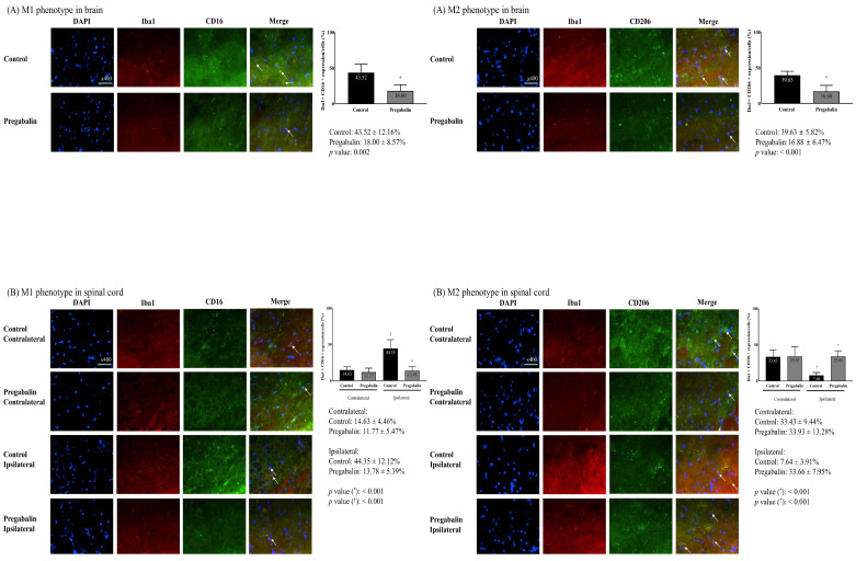
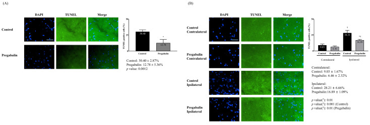
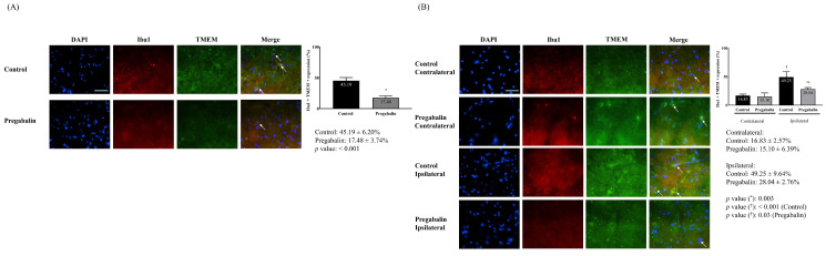
This study investigated the effects of pregabalin on microglial differentiation in rats with neuropathic pain (NP) induced by sciatic nerve ligation and transection. After confirming NP, the rats were randomly allocated to either a pregabalin or control group. The pregabalin group received intraperitoneal injections of 10 mg/kg pregabalin, while the control group received an equivalent volume of normal saline following surgery. On postoperative day 28, neuronal damage, microglial activity, and microglial differentiation were assessed. The pregabalin group exhibited significantly less neuronal damage compared to the control group, along with a significant decrease in activated microglial expression in both the brain and spinal cord. Pregabalin treatment also significantly altered the microglial phenotype expression, with a decrease in the M1 phenotype percentage and an increase in the M2 phenotype percentage in both the brain (M1 phenotype: 43.52 ± 12.16% and 18.00 ± 8.57% in the control and pregabalin groups, respectively; difference: 27.26 [15.18-42.10], p = 0.002; M2 phenotype: 16.88 ± 6.47% and 39.63 ± 5.82% in the control and pregabalin groups, respectively; difference 22.04 [17.17-32.70], p < 0.001) and the spinal cord ipsilateral to nerve injury (M1 phenotype: 44.35 ± 12.12% and 13.78 ± 5.39% in the control and pregabalin groups, respectively; difference 30.46 [21.73-44.45], p < 0.001; M2 phenotype: 7.64 ± 3.91% and 33.66 ± 7.95% in the control and pregabalin groups, respectively; difference 27.41 [21.21-36.30], p < 0.001). Overall, pregabalin treatment significantly decreased the microglial M1 phenotype while increasing the microglial M2 phenotype in NP rats.
本研究探讨了普瑞巴林对坐骨神经结扎和横断诱导的神经病理性疼痛(NP)大鼠小胶质细胞分化的影响。在确认 NP 后,将大鼠随机分配到普瑞巴林组或对照组。普瑞巴林组接受腹腔注射 10mg/kg 普瑞巴林,而对照组在手术后给予等量生理盐水。术后第 28 天,评估神经元损伤、小胶质细胞活性和小胶质细胞分化。与对照组相比,普瑞巴林组的神经元损伤明显减少,大脑和脊髓中活化的小胶质细胞表达也明显减少。普瑞巴林治疗还显著改变了小胶质细胞表型表达,大脑中 M1 表型的百分比降低,M2 表型的百分比增加(M1 表型:对照组和普瑞巴林组分别为 43.52±12.16%和 18.00±8.57%;差异:27.26[15.18-42.10],p=0.002;M2 表型:对照组和普瑞巴林组分别为 16.88±6.47%和 39.63±5.82%;差异 22.04[17.17-32.70],p<0.001),对侧神经损伤脊髓中 M1 表型的百分比降低,M2 表型的百分比增加(M1 表型:对照组和普瑞巴林组分别为 44.35±12.12%和 13.78±5.39%;差异 30.46[21.73-44.45],p<0.001;M2 表型:对照组和普瑞巴林组分别为 7.64±3.91%和 33.66±7.95%;差异 27.41[21.21-36.30],p<0.001)。总之,普瑞巴林治疗显著降低了 NP 大鼠小胶质细胞的 M1 表型,同时增加了小胶质细胞的 M2 表型。